9Q8X | pdb_00009q8x

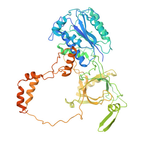
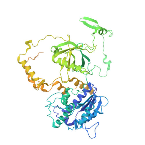
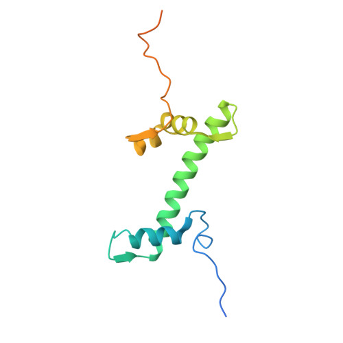
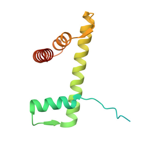
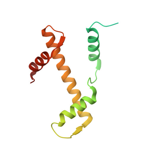
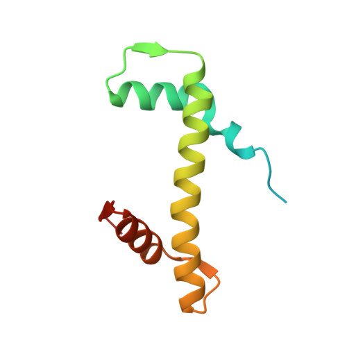
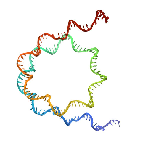
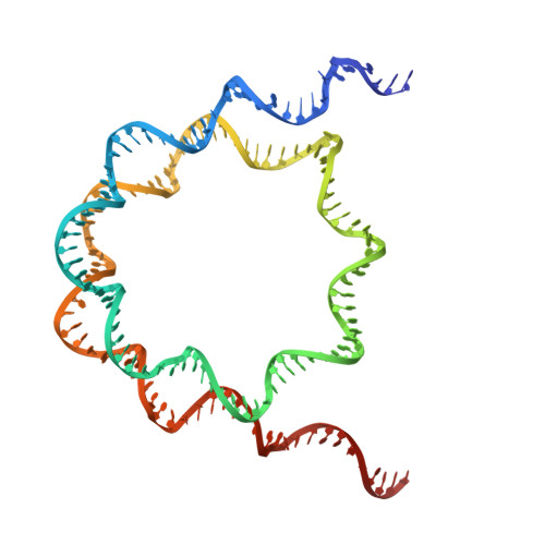
Ku70/80 bound to a 153 bp H2AX nucleosome


Experimental Data Snapshot

  • Method: ELECTRON MICROSCOPY
  • Resolution: 2.94 Å
  • Aggregation State: PARTICLE 
  • Reconstruction Method: SINGLE PARTICLE 

wwPDB Validation 3D Report Full Report

Validation slider image for 9Q8X

This is version 1.1 of the entry. See complete history. 

Literature

Cryo-EM structures of NHEJ assemblies with nucleosomes.

Hall, C., Frit, P., Kefala-Stavridi, A., Pelletier, A., Hardwick, S.W., Amin, H., Bilyard, M.K., Maia De Oliviera, T., Tariq, A., Zahid, S., Chirgadze, D.Y., Balasubramanian, S., Meek, K., Ropars, V., Charbonnier, J.B., Modesti, M., Calsou, P., Britton, S., Blundell, T.L., Schalch, T., Chaplin, A.K.

(2025) Nat Commun 17: 648-648

  • DOI: https://doi.org/10.1038/s41467-025-67376-2
  • Primary Citation Related Structures: 
    9IGW, 9IGX, 9Q80, 9Q8X, 9Q9F, 9QCR, 9QCS, 9QMS

  • PubMed Abstract: 

    DNA double-strand breaks (DSBs) are highly deleterious lesions that can trigger cell death or carcinogenesis if unrepaired or misrepaired. In mammals, most DSBs are repaired by non-homologous end joining (NHEJ), which begins when Ku70/80 binds DNA ends and recruits DNA-PKcs to form the DNA-PK holoenzyme. Although recent cryo-EM studies have resolved several NHEJ assemblies, how these factors access DSBs within nucleosomes remains unclear. Here, we present cryo-EM structures of human Ku70/80 and DNA-PK bound to nucleosomes. Ku70/80 binds the DNA end and bends it away from the nucleosome core, while the Ku70 C-terminal SAP domain makes an additional, specific DNA contact. Our DNA-PK-nucleosome structure further reveals the opening of the Ku80 vWA domain, and we show that non-hydrolysable ATP promotes synapsis by stabilising the Ku80-mediated DNA-PK dimer. These structures reveal a model for DSB recognition on nucleosomal DNA and provide insights relevant to targeting NHEJ in cancer therapy.


  • Organizational Affiliation: 
    • Leicester Institute for Structural and Chemical Biology, Department of Molecular and Cell Biology, University of Leicester, Leicester, UK.

Macromolecule Content 

  • Total Structure Weight: 511.85 kDa 
  • Atom Count: 27,507 
  • Modeled Residue Count: 3,097 
  • Deposited Residue Count: 4,004 
  • Unique protein chains: 6
  • Unique nucleic acid chains: 2

Macromolecules


Find similar proteins by:|  3D Structure
Entity ID: 1
MoleculeChains  Sequence LengthOrganismDetailsImage
X-ray repair cross-complementing protein 6A [auth K],
I [auth M]
609Homo sapiensMutation(s): 0 
Gene Names: XRCC6, G22P1
EC: 3.6.4 (PDB Primary Data), 4.2.99 (PDB Primary Data), 5.6.2.4 (UniProt), 4.2.99.18 (UniProt)
UniProt & NIH Common Fund Data Resources
Find proteins for P12956 (Homo sapiens)
Explore P12956 
Go to UniProtKB:  P12956
PHAROS:  P12956
GTEx:  ENSG00000196419 
Entity Groups
Sequence Clusters30% Identity50% Identity70% Identity90% Identity95% Identity100% Identity
UniProt GroupP12956
Sequence Annotations
Expand
Reference Sequence
Find similar proteins by:|  3D Structure
Entity ID: 2
MoleculeChains  Sequence LengthOrganismDetailsImage
X-ray repair cross-complementing protein 5B [auth L],
J [auth N]
732Homo sapiensMutation(s): 0 
Gene Names: XRCC5, G22P2
EC: 3.6.4 (PDB Primary Data), 5.6.2.4 (UniProt), 4.2.99.18 (UniProt)
UniProt & NIH Common Fund Data Resources
Find proteins for P13010 (Homo sapiens)
Explore P13010 
Go to UniProtKB:  P13010
PHAROS:  P13010
GTEx:  ENSG00000079246 
Entity Groups
Sequence Clusters30% Identity50% Identity70% Identity90% Identity95% Identity100% Identity
UniProt GroupP13010
Sequence Annotations
Expand
Reference Sequence
Find similar proteins by:|  3D Structure
Entity ID: 3
MoleculeChains  Sequence LengthOrganismDetailsImage
Histone H2AXC [auth G],
G [auth C]
143Homo sapiensMutation(s): 0 
Gene Names: H2AX, H2AFX
UniProt & NIH Common Fund Data Resources
Find proteins for P16104 (Homo sapiens)
Explore P16104 
Go to UniProtKB:  P16104
PHAROS:  P16104
GTEx:  ENSG00000188486 
Entity Groups
Sequence Clusters30% Identity50% Identity70% Identity90% Identity95% Identity100% Identity
UniProt GroupP16104
Sequence Annotations
Expand
Reference Sequence
Find similar proteins by:|  3D Structure
Entity ID: 4
MoleculeChains  Sequence LengthOrganismDetailsImage
Histone H2B type 1-JD [auth H],
H [auth D]
126Homo sapiensMutation(s): 0 
Gene Names: H2BC11, H2BFR, HIST1H2BJ
UniProt & NIH Common Fund Data Resources
Find proteins for P06899 (Homo sapiens)
Explore P06899 
Go to UniProtKB:  P06899
PHAROS:  P06899
GTEx:  ENSG00000124635 
Entity Groups
Sequence Clusters30% Identity50% Identity70% Identity90% Identity95% Identity100% Identity
UniProt GroupP06899
Sequence Annotations
Expand
Reference Sequence
Find similar proteins by:|  3D Structure
Entity ID: 5
MoleculeChains  Sequence LengthOrganismDetailsImage
Histone H3.1E,
K [auth A]
136Homo sapiensMutation(s): 0 
Gene Names: 
UniProt & NIH Common Fund Data Resources
Find proteins for P68431 (Homo sapiens)
Explore P68431 
Go to UniProtKB:  P68431
Entity Groups
Sequence Clusters30% Identity50% Identity70% Identity90% Identity95% Identity100% Identity
UniProt GroupP68431
Sequence Annotations
Expand
Reference Sequence
Find similar proteins by:|  3D Structure
Entity ID: 6
MoleculeChains  Sequence LengthOrganismDetailsImage
Histone H4F [auth B],
L [auth F]
103Homo sapiensMutation(s): 0 
Gene Names: 
UniProt & NIH Common Fund Data Resources
Find proteins for P62805 (Homo sapiens)
Explore P62805 
Go to UniProtKB:  P62805
Entity Groups
Sequence Clusters30% Identity50% Identity70% Identity90% Identity95% Identity100% Identity
UniProt GroupP62805
Sequence Annotations
Expand
Reference Sequence
Find similar nucleic acids by:  (by identity cutoff) 
Entity ID: 7
MoleculeChains LengthOrganismImage
DNAM [auth I]153Homo sapiens
Sequence Annotations
Expand
Reference Sequence
Find similar nucleic acids by:  (by identity cutoff) 
Entity ID: 8
MoleculeChains LengthOrganismImage
DNAN [auth J]153Homo sapiens
Sequence Annotations
Expand
Reference Sequence

Experimental Data & Validation

Experimental Data

  • Method: ELECTRON MICROSCOPY
  • Resolution: 2.94 Å
  • Aggregation State: PARTICLE 
  • Reconstruction Method: SINGLE PARTICLE 
EM Software:
TaskSoftware PackageVersion
RECONSTRUCTIONPHENIX

Structure Validation

View Full Validation Report


Entry History 

& Funding Information

Deposition Data


Funding OrganizationLocationGrant Number
Medical Research Council (MRC, Canada)CanadaMR/X00029X/1
The Lister Institute of Preventive MedicineUnited Kingdom--

Revision History  (Full details and data files)

  • Version 1.0: 2026-01-14
    Type: Initial release
  • Version 1.1: 2026-01-28
    Changes: Data collection, Database references