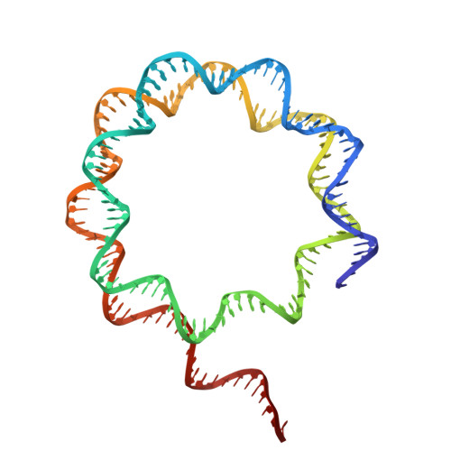
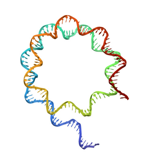
8VMJ | pdb_00008vmj

H3K4me3 nucleosome bound to PRC2_AJ119-450


Experimental Data Snapshot

  • Method: ELECTRON MICROSCOPY
  • Resolution: 3.10 Å
  • Aggregation State: PARTICLE 
  • Reconstruction Method: SINGLE PARTICLE 

wwPDB Validation   3D Report Full Report


This is version 1.2 of the entry. See complete history


Literature

Structural basis for the inhibition of PRC2 by active transcription histone posttranslational modifications.

Cookis, T.Lydecker, A.Sauer, P.Kasinath, V.Nogales, E.

(2025) Nat Struct Mol Biol 32: 393-404

  • DOI: https://doi.org/10.1038/s41594-024-01452-x
  • Primary Citation of Related Structures:  
    8VMI, 8VMJ, 8VML, 8VMN, 8VNV, 8VNZ, 8VO0, 8VOB

  • PubMed Abstract: 

    Polycomb repressive complex 2 (PRC2) trimethylates histone H3 on K27 (H3K27me3) leading to gene silencing that is essential for embryonic development and maintenance of cell identity. PRC2 is regulated by protein cofactors and their crosstalk with histone modifications. Trimethylated histone H3 on K4 (H3K4me3) and K36 (H3K36me3) localize to sites of active transcription and inhibit PRC2 activity through unknown mechanisms. Using cryo-electron microscopy, we reveal that histone H3 tails containing H3K36me3 engage poorly with PRC2 and preclude its effective interaction with chromatin, while H3K4me3 binds to the allosteric site in the EED subunit, acting as an antagonist that competes with activators required for spreading of the H3K27me3 repressive mark. Thus, the location of the H3K4me3 and H3K36me3 modifications along the H3 tail allows them to target two requirements for efficient trimethylation of H3K27 by PRC2. We further show that the JARID2 cofactor modulates PRC2 activity in the presence of these histone modifications.


  • Organizational Affiliation

    Department of Molecular and Cell Biology, University of California, Berkeley, CA, USA.


Macromolecules

Find similar proteins by:  (by identity cutoff)  |  3D Structure
Entity ID: 2
MoleculeChains Sequence LengthOrganismDetailsImage
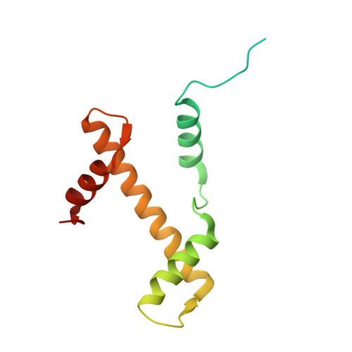
Histone H3.2B [auth I],
F [auth O]
136Homo sapiensMutation(s): 0 
Gene Names: H3C15HIST2H3AH3C14H3F2H3FMHIST2H3CH3C13HIST2H3D
UniProt & NIH Common Fund Data Resources
Find proteins for Q71DI3 (Homo sapiens)
Explore Q71DI3 
Go to UniProtKB:  Q71DI3
PHAROS:  Q71DI3
Entity Groups  
Sequence Clusters30% Identity50% Identity70% Identity90% Identity95% Identity100% Identity
UniProt GroupQ71DI3
Sequence Annotations
Expand
  • Reference Sequence
Find similar proteins by:  (by identity cutoff)  |  3D Structure
Entity ID: 3
MoleculeChains Sequence LengthOrganismDetailsImage
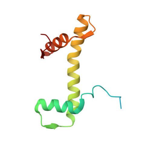
Histone H4C [auth J],
G [auth Q]
103Homo sapiensMutation(s): 0 
Gene Names: 
UniProt & NIH Common Fund Data Resources
Find proteins for P62805 (Homo sapiens)
Explore P62805 
Go to UniProtKB:  P62805
PHAROS:  P62805
Entity Groups  
Sequence Clusters30% Identity50% Identity70% Identity90% Identity95% Identity100% Identity
UniProt GroupP62805
Sequence Annotations
Expand
  • Reference Sequence
Find similar proteins by:  (by identity cutoff)  |  3D Structure
Entity ID: 4
MoleculeChains Sequence LengthOrganismDetailsImage
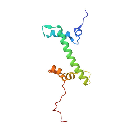
Histone H2AD [auth K],
H [auth R]
108Xenopus laevisMutation(s): 0 
Gene Names: LOC494591h2ac14.Lhist1h2ajhist1h2aj.L
UniProt
Find proteins for Q6AZJ8 (Xenopus laevis)
Explore Q6AZJ8 
Go to UniProtKB:  Q6AZJ8
Entity Groups  
Sequence Clusters30% Identity50% Identity70% Identity90% Identity95% Identity100% Identity
UniProt GroupQ6AZJ8
Sequence Annotations
Expand
  • Reference Sequence
Find similar proteins by:  (by identity cutoff)  |  3D Structure
Entity ID: 5
MoleculeChains Sequence LengthOrganismDetailsImage
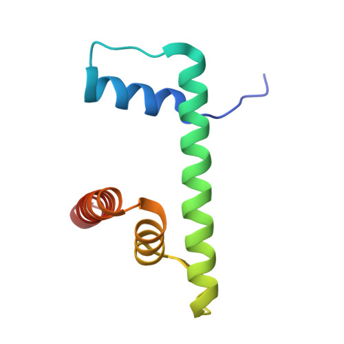
Histone H2BE [auth M],
I [auth S]
96Xenopus laevisMutation(s): 0 
Gene Names: LOC108704302
UniProt
Find proteins for A0A8J1LZU9 (Xenopus laevis)
Explore A0A8J1LZU9 
Go to UniProtKB:  A0A8J1LZU9
Entity Groups  
Sequence Clusters30% Identity50% Identity70% Identity90% Identity95% Identity100% Identity
UniProt GroupA0A8J1LZU9
Sequence Annotations
Expand
  • Reference Sequence
Small Molecules
Modified Residues  1 Unique
IDChains TypeFormula2D DiagramParent
M3L
Query on M3L
B [auth I],
F [auth O]
L-PEPTIDE LINKINGC9 H21 N2 O2LYS
Experimental Data & Validation

Experimental Data

  • Method: ELECTRON MICROSCOPY
  • Resolution: 3.10 Å
  • Aggregation State: PARTICLE 
  • Reconstruction Method: SINGLE PARTICLE 
EM Software:
TaskSoftware PackageVersion
MODEL REFINEMENTPHENIX1.20.1_4487

Structure Validation

View Full Validation Report


Entry History & Funding Information

Deposition Data


Funding OrganizationLocationGrant Number
National Institutes of Health/National Institute of General Medical Sciences (NIH/NIGMS)United StatesR35-GM127018
National Science Foundation (NSF, United States)United StatesGM-08295

Revision History  (Full details and data files)

  • Version 1.0: 2025-01-15
    Type: Initial release
  • Version 1.1: 2025-01-22
    Changes: Data collection, Database references
  • Version 1.2: 2025-03-05
    Changes: Data collection, Database references