8AAG

H1-bound palindromic nucleosome, state 1


Experimental Data Snapshot

  • Method: ELECTRON MICROSCOPY
  • Resolution: 10.0 Å
  • Aggregation State: PARTICLE 
  • Reconstruction Method: SINGLE PARTICLE 

Starting Model: experimental
View more details

wwPDB Validation   3D Report Full Report


This is version 1.3 of the entry. See complete history


Literature

Nucleosome dyad determines the H1 C-terminus collapse on distinct DNA arms.

Louro, J.A.Boopathi, R.Beinsteiner, B.Mohideen Patel, A.K.Cheng, T.C.Angelov, D.Hamiche, A.Bendar, J.Kale, S.Klaholz, B.P.Dimitrov, S.

(2023) Structure 31: 201

  • DOI: https://doi.org/10.1016/j.str.2022.12.005
  • Primary Citation of Related Structures:  
    8AAG

  • PubMed Abstract: 

    Nucleosomes are symmetric structures. However, binding of linker histones generates an inherently asymmetric H1-nucleosome complex, and whether this asymmetry is transmitted to the overall nucleosome structure, and therefore also to chromatin, is unclear. Efforts to investigate potential asymmetry due to H1s have been hampered by the DNA sequence, which naturally differs in each gyre. To overcome this issue, we designed and analyzed by cryo-EM a nucleosome reconstituted with a palindromic (601L) 197-bp DNA. As in the non-palindromic 601 sequence, H1 restricts linker DNA flexibility but reveals partial asymmetrical unwrapping. However, in contrast to the non-palindromic nucleosome, in the palindromic nucleosome H1 CTD collapses to the proximal linker. Molecular dynamics simulations show that this could be dictated by a slightly tilted orientation of the globular domain (GD) of H1, which could be linked to the DNA sequence of the nucleosome dyad.


  • Organizational Affiliation

    Centre for Integrative Biology (CBI), Department of Integrated Structural Biology, IGBMC (Institute of Genetics and of Molecular and Cellular Biology), 1 rue Laurent Fries, 67404 Illkirch, France.


Macromolecules

Find similar proteins by:  (by identity cutoff)  |  3D Structure
Entity ID: 3
MoleculeChains Sequence LengthOrganismDetailsImage
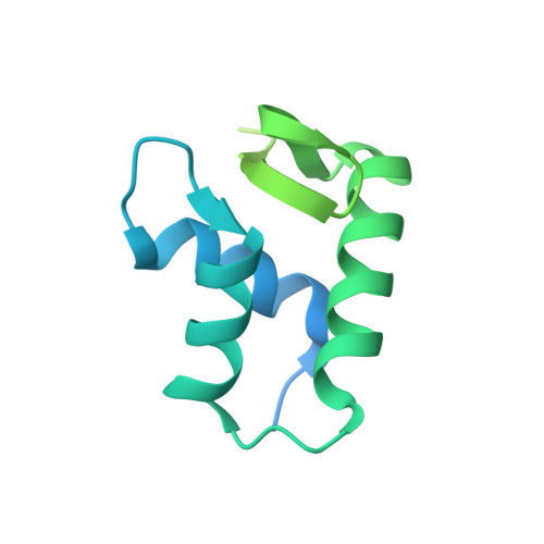
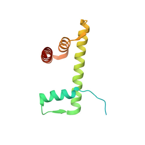
Histone H1.0-BC [auth Z]196Xenopus laevisMutation(s): 0 
Gene Names: h1f0-b
UniProt
Find proteins for P22844 (Xenopus laevis)
Go to UniProtKB:  P22844
Sequence Annotations
Expand
  • Reference Sequence
Find similar proteins by:  (by identity cutoff)  |  3D Structure
Entity ID: 4
MoleculeChains Sequence LengthOrganismDetailsImage
Histone H3.2D [auth A],
H [auth E]
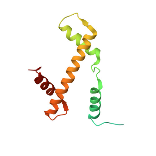
136Homo sapiensMutation(s): 0 
UniProt & NIH Common Fund Data Resources
Find proteins for Q71DI3 (Homo sapiens)
Explore Q71DI3 
Go to UniProtKB:  Q71DI3
PHAROS:  Q71DI3
Entity Groups  
Sequence Clusters30% Identity50% Identity70% Identity90% Identity95% Identity100% Identity
UniProt GroupQ71DI3
Sequence Annotations
Expand
  • Reference Sequence
Find similar proteins by:  (by identity cutoff)  |  3D Structure
Entity ID: 5
MoleculeChains Sequence LengthOrganismDetailsImage
Histone H4E [auth B],
I [auth F]
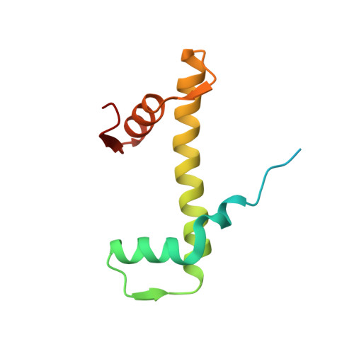
103Homo sapiensMutation(s): 0 
Gene Names: 
UniProt & NIH Common Fund Data Resources
Find proteins for P62805 (Homo sapiens)
Go to UniProtKB:  P62805
PHAROS:  P62805
Sequence Annotations
Expand
  • Reference Sequence
Find similar proteins by:  (by identity cutoff)  |  3D Structure
Entity ID: 6
MoleculeChains Sequence LengthOrganismDetailsImage
Histone H2A type 1F [auth C],
J [auth G]
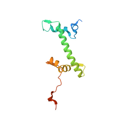
129Homo sapiensMutation(s): 0 
UniProt
Find proteins for P06897 (Xenopus laevis)
Go to UniProtKB:  P06897
Sequence Annotations
Expand
  • Reference Sequence
Find similar proteins by:  (by identity cutoff)  |  3D Structure
Entity ID: 7
MoleculeChains Sequence LengthOrganismDetailsImage
Histone H2B type 1-C/E/F/G/IG [auth D],
K [auth H]
122Homo sapiensMutation(s): 0 
UniProt
Find proteins for P02281 (Xenopus laevis)
Go to UniProtKB:  P02281
Sequence Annotations
Expand
  • Reference Sequence
Find similar nucleic acids by: 3D Structure
Entity ID: 1
MoleculeChains LengthOrganismImage
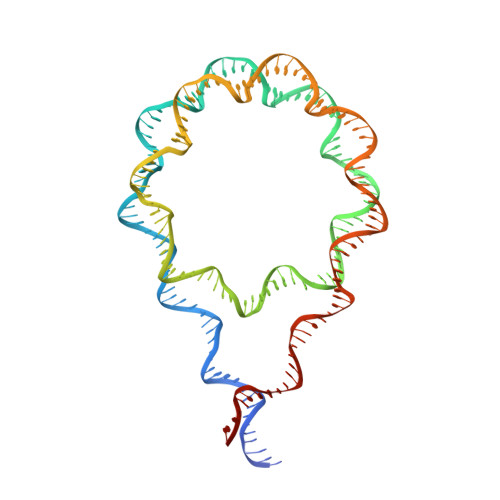
DNA/RNA (185-MER)A [auth I]198synthetic construct
Sequence Annotations
Expand
  • Reference Sequence
Find similar nucleic acids by: 3D Structure
Entity ID: 2
MoleculeChains LengthOrganismImage
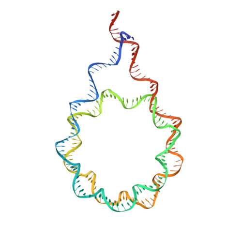
DNA/RNA (185-MER)B [auth J]197synthetic construct
Sequence Annotations
Expand
  • Reference Sequence
Experimental Data & Validation

Experimental Data

  • Method: ELECTRON MICROSCOPY
  • Resolution: 10.0 Å
  • Aggregation State: PARTICLE 
  • Reconstruction Method: SINGLE PARTICLE 
EM Software:
TaskSoftware PackageVersion
RECONSTRUCTIONRELION3.0
MODEL REFINEMENTPHENIX1.18

Structure Validation

View Full Validation Report


Entry History & Funding Information

Deposition Data


Funding OrganizationLocationGrant Number
Centre National de la Recherche Scientifique (CNRS)France--
Agence Nationale de la Recherche (ANR)France--
French Infrastructure for Integrated Structural Biology (FRISBI)France--
Instruct-ERIC Center (Strasbourg Centre)France--
Foundation for Medical Research (France)France--
Institut National du Cancer (inCA)--
European Regional Development FundEuropean UnionepiRNA - (Region Grand Est; Competitivite Alsace 2014-2020)
European Molecular Biology Organization (EMBO)European UnionEMBO IG 5056

Revision History  (Full details and data files)

  • Version 1.0: 2022-12-14
    Type: Initial release
  • Version 1.1: 2023-01-18
    Changes: Database references
  • Version 1.2: 2023-02-15
    Changes: Database references
  • Version 1.3: 2024-07-24
    Changes: Data collection, Refinement description