5JRG

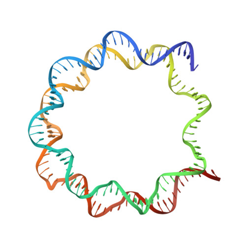
Crystal structure of the nucleosome containing the DNA with tetrahydrofuran (THF)


Experimental Data Snapshot

  • Method: X-RAY DIFFRACTION
  • Resolution: 2.50 Å
  • R-Value Free: 0.245 
  • R-Value Work: 0.193 
  • R-Value Observed: 0.196 

Starting Model: experimental
View more details

wwPDB Validation   3D Report Full Report


This is version 1.2 of the entry. See complete history


Literature

Polymorphism of apyrimidinic DNA structures in the nucleosome

Osakabe, A.Arimura, Y.Matsumoto, S.Horikoshi, N.Sugasawa, K.Kurumizaka, H.

(2017) Sci Rep 7: 41783-41783

  • DOI: https://doi.org/10.1038/srep41783
  • Primary Citation of Related Structures:  
    5JRG

  • PubMed Abstract: 

    Huge amounts (>10,000/day) of apurinic/apyrimidinic (AP) sites are produced in genomes, but their structures in chromatin remain undetermined. We determined the crystal structure of the nucleosome containing AP-site analogs at two symmetric sites, which revealed structural polymorphism: one forms an inchworm configuration without an empty space at the AP site, and the other forms a B-form-like structure with an empty space and the orphan base. This unexpected inchworm configuration of the AP site is important to understand the AP DNA repair mechanism, because it may not be recognized by the major AP-binding protein, APE1, during the base excision repair process.


  • Organizational Affiliation

    Laboratory of Structural Biology, Graduate School of Advanced Science and Engineering, Waseda University, 2-2 Wakamatsu-cho, Shinjuku-ku, Tokyo 162-8480, Japan.


Macromolecules

Find similar proteins by:  (by identity cutoff)  |  3D Structure
Entity ID: 1
MoleculeChains Sequence LengthOrganismDetailsImage
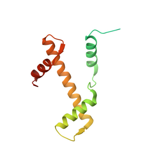
Histone H3.1
A, E
139Homo sapiensMutation(s): 0 
Gene Names: 
UniProt & NIH Common Fund Data Resources
Find proteins for P68431 (Homo sapiens)
Explore P68431 
Go to UniProtKB:  P68431
PHAROS:  P68431
Entity Groups  
Sequence Clusters30% Identity50% Identity70% Identity90% Identity95% Identity100% Identity
UniProt GroupP68431
Sequence Annotations
Expand
  • Reference Sequence
Find similar proteins by:  (by identity cutoff)  |  3D Structure
Entity ID: 2
MoleculeChains Sequence LengthOrganismDetailsImage
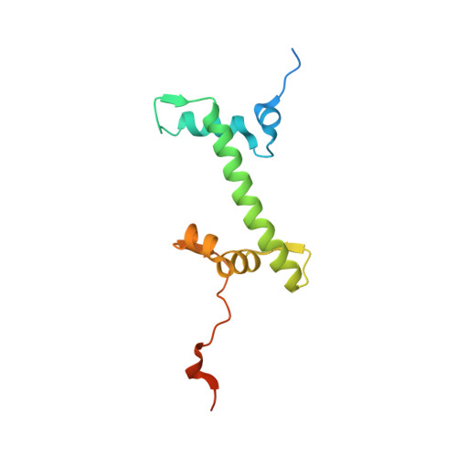
Histone H4
B, F
106Homo sapiensMutation(s): 0 
Gene Names: 
UniProt & NIH Common Fund Data Resources
Find proteins for P62805 (Homo sapiens)
Explore P62805 
Go to UniProtKB:  P62805
PHAROS:  P62805
Entity Groups  
Sequence Clusters30% Identity50% Identity70% Identity90% Identity95% Identity100% Identity
UniProt GroupP62805
Sequence Annotations
Expand
  • Reference Sequence
Find similar proteins by:  (by identity cutoff)  |  3D Structure
Entity ID: 3
MoleculeChains Sequence LengthOrganismDetailsImage
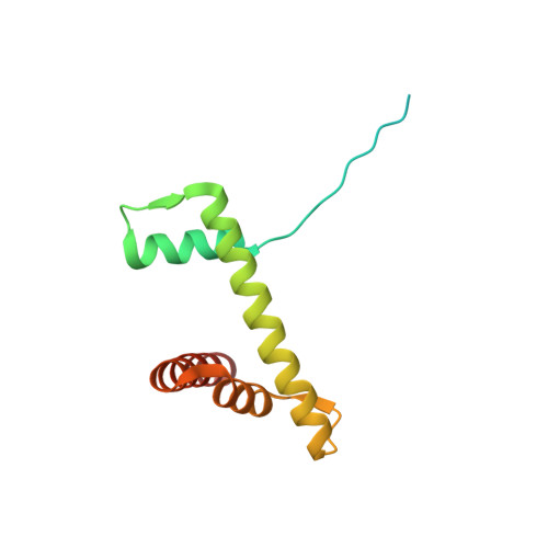
Histone H2A type 1-B/E
C, G
133Homo sapiensMutation(s): 0 
Gene Names: HIST1H2ABH2AFMHIST1H2AEH2AFA
UniProt & NIH Common Fund Data Resources
Find proteins for P04908 (Homo sapiens)
Explore P04908 
Go to UniProtKB:  P04908
PHAROS:  P04908
Entity Groups  
Sequence Clusters30% Identity50% Identity70% Identity90% Identity95% Identity100% Identity
UniProt GroupP04908
Sequence Annotations
Expand
  • Reference Sequence
Find similar proteins by:  (by identity cutoff)  |  3D Structure
Entity ID: 4
MoleculeChains Sequence LengthOrganismDetailsImage
Histone H2B type 1-J
D, H
129Homo sapiensMutation(s): 0 
Gene Names: HIST1H2BJH2BFR
UniProt & NIH Common Fund Data Resources
Find proteins for P06899 (Homo sapiens)
Explore P06899 
Go to UniProtKB:  P06899
PHAROS:  P06899
GTEx:  ENSG00000124635 
Entity Groups  
Sequence Clusters30% Identity50% Identity70% Identity90% Identity95% Identity100% Identity
UniProt GroupP06899
Sequence Annotations
Expand
  • Reference Sequence
Find similar nucleic acids by:  (by identity cutoff)  |  3D Structure
Entity ID: 5
MoleculeChains LengthOrganismImage
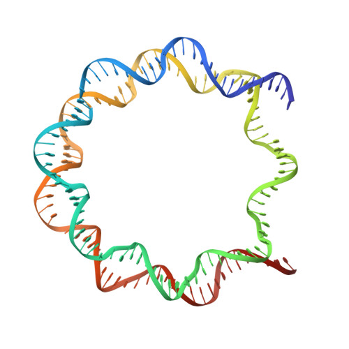
DNA (145-MER)145Homo sapiens
Sequence Annotations
Expand
  • Reference Sequence
Find similar nucleic acids by:  (by identity cutoff)  |  3D Structure
Entity ID: 6
MoleculeChains LengthOrganismImage
DNA (145-MER)145Homo sapiens
Sequence Annotations
Expand
  • Reference Sequence
Small Molecules
Ligands 2 Unique
IDChains Name / Formula / InChI Key2D Diagram3D Interactions
MN
Query on MN

Download Ideal Coordinates CCD File 
AA [auth J]
BA [auth J]
CA [auth J]
M [auth E]
P [auth I]
AA [auth J],
BA [auth J],
CA [auth J],
M [auth E],
P [auth I],
Q [auth I],
R [auth I],
S [auth I],
T [auth I],
U [auth I],
V [auth I],
W [auth I],
X [auth J],
Y [auth J],
Z [auth J]
MANGANESE (II) ION
Mn
WAEMQWOKJMHJLA-UHFFFAOYSA-N
CL
Query on CL

Download Ideal Coordinates CCD File 
K [auth A],
L [auth C],
N [auth E],
O [auth G]
CHLORIDE ION
Cl
VEXZGXHMUGYJMC-UHFFFAOYSA-M
Experimental Data & Validation

Experimental Data

  • Method: X-RAY DIFFRACTION
  • Resolution: 2.50 Å
  • R-Value Free: 0.245 
  • R-Value Work: 0.193 
  • R-Value Observed: 0.196 
  • Space Group: P 21 21 21
Unit Cell:
Length ( Å )Angle ( ˚ )
a = 99.491α = 90
b = 108.773β = 90
c = 169.571γ = 90
Software Package:
Software NamePurpose
HKL-2000data scaling
PHENIXrefinement
PDB_EXTRACTdata extraction
Cootmodel building
PHASERphasing

Structure Validation

View Full Validation Report


Entry History & Funding Information

Deposition Data


Funding OrganizationLocationGrant Number
MEXT KAKENHIJapan25116002
JSPS KAKENHIJapan25250023
Platform Project for Supporting Drug Discovery and Life Science ResearchJapan--
JSPS KAKENHIJapan15H06691

Revision History  (Full details and data files)

  • Version 1.0: 2017-03-08
    Type: Initial release
  • Version 1.1: 2017-10-04
    Changes: Data collection
  • Version 1.2: 2023-11-08
    Changes: Data collection, Database references, Derived calculations, Refinement description