6UB7

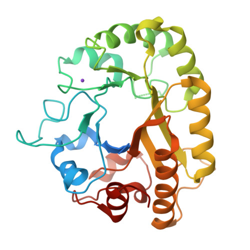
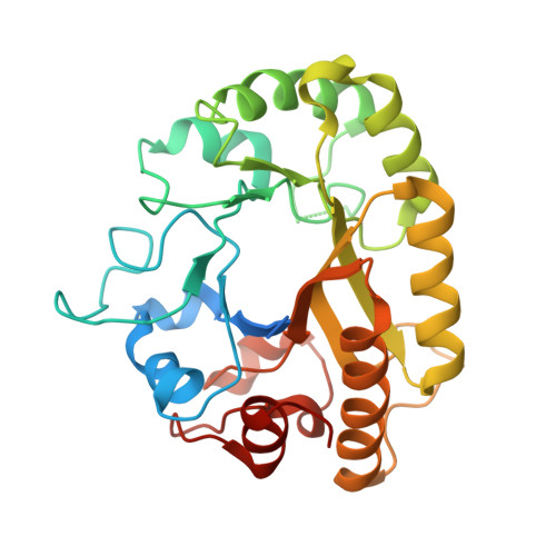
Crystal structure of a GH128 (subgroup V) exo-beta-1,3-glucanase from Cryptococcus neoformans (CnGH128_V)


Experimental Data Snapshot

  • Method: X-RAY DIFFRACTION
  • Resolution: 1.80 Å
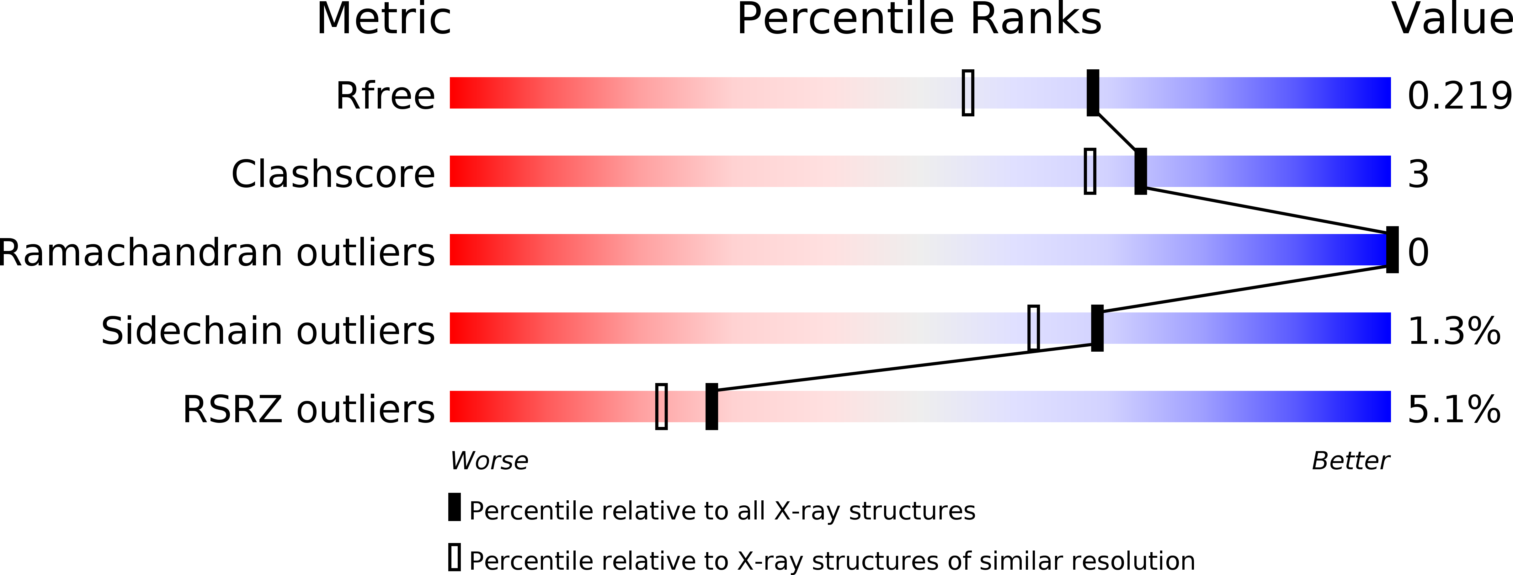
  • R-Value Free: 0.218 
  • R-Value Work: 0.185 
  • R-Value Observed: 0.186 

wwPDB Validation   3D Report Full Report


This is version 1.3 of the entry. See complete history


Literature

Structural insights into beta-1,3-glucan cleavage by a glycoside hydrolase family.

Santos, C.R.Costa, P.A.C.R.Vieira, P.S.Gonzalez, S.E.T.Correa, T.L.R.Lima, E.A.Mandelli, F.Pirolla, R.A.S.Domingues, M.N.Cabral, L.Martins, M.P.Cordeiro, R.L.Junior, A.T.Souza, B.P.Prates, E.T.Gozzo, F.C.Persinoti, G.F.Skaf, M.S.Murakami, M.T.

(2020) Nat Chem Biol 16: 920-929

  • DOI: https://doi.org/10.1038/s41589-020-0554-5
  • Primary Citation of Related Structures:  
    6UAQ, 6UAR, 6UAS, 6UAT, 6UAU, 6UAV, 6UAW, 6UAX, 6UAY, 6UAZ, 6UB0, 6UB1, 6UB2, 6UB3, 6UB4, 6UB5, 6UB6, 6UB7, 6UB8, 6UBA, 6UBB, 6UBC, 6UBD, 6UFL, 6UFZ

  • PubMed Abstract: 

    The fundamental and assorted roles of β-1,3-glucans in nature are underpinned on diverse chemistry and molecular structures, demanding sophisticated and intricate enzymatic systems for their processing. In this work, the selectivity and modes of action of a glycoside hydrolase family active on β-1,3-glucans were systematically investigated combining sequence similarity network, phylogeny, X-ray crystallography, enzyme kinetics, mutagenesis and molecular dynamics. This family exhibits a minimalist and versatile (α/β)-barrel scaffold, which can harbor distinguishing exo or endo modes of action, including an ancillary-binding site for the anchoring of triple-helical β-1,3-glucans. The substrate binding occurs via a hydrophobic knuckle complementary to the canonical curved conformation of β-1,3-glucans or through a substrate conformational change imposed by the active-site topology of some fungal enzymes. Together, these findings expand our understanding of the enzymatic arsenal of bacteria and fungi for the breakdown and modification of β-1,3-glucans, which can be exploited for biotechnological applications.


  • Organizational Affiliation

    Brazilian Biorenewables National Laboratory, Brazilian Center for Research in Energy and Materials, Campinas, São Paulo, Brazil.


Macromolecules
Find similar proteins by:  (by identity cutoff)  |  3D Structure
Entity ID: 1
MoleculeChains Sequence LengthOrganismDetailsImage
Glyco_hydro_cc domain-containing protein287Cryptococcus neoformansMutation(s): 0 
UniProt
Find proteins for J9VQQ4 (Cryptococcus neoformans var. grubii serotype A (strain H99 / ATCC 208821 / CBS 10515 / FGSC 9487))
Explore J9VQQ4 
Go to UniProtKB:  J9VQQ4
Entity Groups  
Sequence Clusters30% Identity50% Identity70% Identity90% Identity95% Identity100% Identity
UniProt GroupJ9VQQ4
Sequence Annotations
Expand
  • Reference Sequence
Small Molecules
Ligands 1 Unique
IDChains Name / Formula / InChI Key2D Diagram3D Interactions
K
Query on K

Download Ideal Coordinates CCD File 
B [auth A]POTASSIUM ION
K
NPYPAHLBTDXSSS-UHFFFAOYSA-N
Experimental Data & Validation

Experimental Data

  • Method: X-RAY DIFFRACTION
  • Resolution: 1.80 Å
  • R-Value Free: 0.218 
  • R-Value Work: 0.185 
  • R-Value Observed: 0.186 
  • Space Group: P 21 21 21
Unit Cell:
Length ( Å )Angle ( ˚ )
a = 58.896α = 90
b = 60.066β = 90
c = 96.085γ = 90
Software Package:
Software NamePurpose
REFMACrefinement
PDB_EXTRACTdata extraction
XDSdata reduction
XSCALEdata scaling
PHASERphasing

Structure Validation

View Full Validation Report


Entry History & Funding Information

Deposition Data


Funding OrganizationLocationGrant Number
Sao Paulo Research Foundation (FAPESP)Brazil15/26982-0

Revision History  (Full details and data files)

  • Version 1.0: 2020-05-20
    Type: Initial release
  • Version 1.1: 2020-06-10
    Changes: Database references
  • Version 1.2: 2020-08-05
    Changes: Database references
  • Version 1.3: 2024-03-13
    Changes: Data collection, Database references