6UB6 | pdb_00006ub6

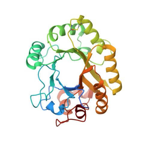
Crystal structure of a GH128 (subgroup IV) endo-beta-1,3-glucanase from Lentinula edodes (LeGH128_IV) in complex with laminaritetraose


Experimental Data Snapshot

  • Method: X-RAY DIFFRACTION
  • Resolution: 1.25 Å
  • R-Value Free: 
    0.191 (Depositor), 0.199 (DCC) 
  • R-Value Work: 
    0.174 (Depositor), 0.183 (DCC) 

wwPDB Validation 3D Report Full Report

Validation slider image for 6UB6

This is version 2.2 of the entry. See complete history. 

Literature

Structural insights into beta-1,3-glucan cleavage by a glycoside hydrolase family.

Santos, C.R., Costa, P.A.C.R., Vieira, P.S., Gonzalez, S.E.T., Correa, T.L.R., Lima, E.A., Mandelli, F., Pirolla, R.A.S., Domingues, M.N., Cabral, L., Martins, M.P., Cordeiro, R.L., Junior, A.T., Souza, B.P., Prates, E.T., Gozzo, F.C., Persinoti, G.F., Skaf, M.S., Murakami, M.T.

(2020) Nat Chem Biol 16: 920-929

  • DOI: https://doi.org/10.1038/s41589-020-0554-5
  • Primary Citation Related Structures: 
    6UAQ, 6UAR, 6UAS, 6UAT, 6UAU, 6UAV, 6UAW, 6UAX, 6UAY, 6UAZ, 6UB0, 6UB1, 6UB2, 6UB3, 6UB4, 6UB5, 6UB6, 6UB7, 6UB8, 6UBA, 6UBB, 6UBC, 6UBD, 6UFL, 6UFZ

  • PubMed Abstract: 

    The fundamental and assorted roles of β-1,3-glucans in nature are underpinned on diverse chemistry and molecular structures, demanding sophisticated and intricate enzymatic systems for their processing. In this work, the selectivity and modes of action of a glycoside hydrolase family active on β-1,3-glucans were systematically investigated combining sequence similarity network, phylogeny, X-ray crystallography, enzyme kinetics, mutagenesis and molecular dynamics. This family exhibits a minimalist and versatile (α/β)-barrel scaffold, which can harbor distinguishing exo or endo modes of action, including an ancillary-binding site for the anchoring of triple-helical β-1,3-glucans. The substrate binding occurs via a hydrophobic knuckle complementary to the canonical curved conformation of β-1,3-glucans or through a substrate conformational change imposed by the active-site topology of some fungal enzymes. Together, these findings expand our understanding of the enzymatic arsenal of bacteria and fungi for the breakdown and modification of β-1,3-glucans, which can be exploited for biotechnological applications.


  • Organizational Affiliation: 
    • Brazilian Biorenewables National Laboratory, Brazilian Center for Research in Energy and Materials, Campinas, São Paulo, Brazil.

Macromolecule Content 

  • Total Structure Weight: 29.83 kDa 
  • Atom Count: 2,358 
  • Modeled Residue Count: 252 
  • Deposited Residue Count: 270 
  • Unique protein chains: 1

Macromolecules

Find similar proteins by:|  3D Structure
Entity ID: 1
MoleculeChains  Sequence LengthOrganismDetailsImage
Endo-beta-1,3-glucanase270Lentinula edodesMutation(s): 0 
Gene Names: glu1
UniProt
Find proteins for G9M5R4 (Lentinula edodes)
Explore G9M5R4 
Go to UniProtKB:  G9M5R4
Entity Groups
Sequence Clusters30% Identity50% Identity70% Identity90% Identity95% Identity100% Identity
UniProt GroupG9M5R4
Sequence Annotations
Expand
Reference Sequence

Oligosaccharides

Help  
Entity ID: 2
MoleculeChains Length2D Diagram GlycosylationD Interactions
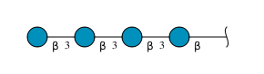
beta-D-glucopyranose-(1-3)-beta-D-glucopyranose-(1-3)-beta-D-glucopyranose-(1-3)-beta-D-glucopyranose
B
4N/A
Glycosylation Resources
GlyTouCan: G87841AX
GlyCosmos: G87841AX
GlyGen: G87841AX
Entity ID: 3
MoleculeChains Length2D Diagram GlycosylationD Interactions
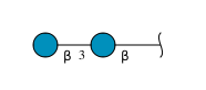
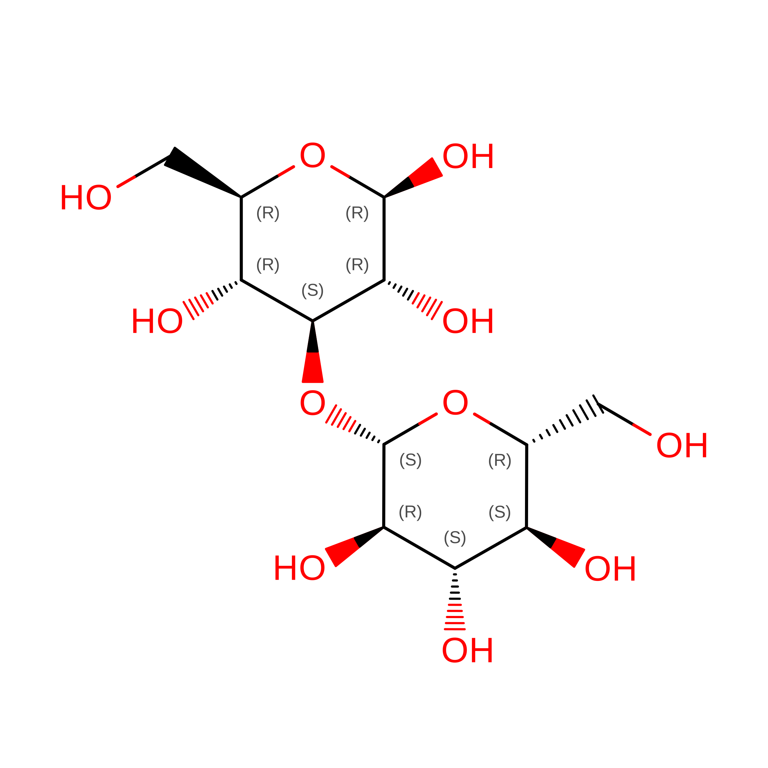
beta-D-glucopyranose-(1-3)-beta-D-glucopyranose
C
2N/A
Glycosylation Resources
GlyTouCan: G36535HU
GlyCosmos: G36535HU

Small Molecules

Ligands 1 Unique
IDChains Name / Formula / InChI Key2D Diagram3D Interactions
CL

Query on CL


Download:Ideal Coordinates CCD File
D [auth A]CHLORIDE ION
Cl
VEXZGXHMUGYJMC-UHFFFAOYSA-M

Biologically Interesting Molecules (External Reference) 

1 Unique

Experimental Data & Validation

Experimental Data

  • Method: X-RAY DIFFRACTION
  • Resolution: 1.25 Å
  • R-Value Free:  0.191 (Depositor), 0.199 (DCC) 
  • R-Value Work:  0.174 (Depositor), 0.183 (DCC) 
Space Group: P 1 21 1
Unit Cell:
Length ( Å )Angle ( ˚ )
a = 45.871α = 90
b = 47.678β = 110.42
c = 52.549γ = 90
Software Package:
Software NamePurpose
REFMACrefinement
XDSdata reduction
XSCALEdata scaling
MOLREPphasing

Structure Validation

View Full Validation Report


Entry History 

& Funding Information

Deposition Data


Funding OrganizationLocationGrant Number
Sao Paulo Research Foundation (FAPESP)Brazil15/26982-0

Revision History  (Full details and data files)

  • Version 1.0: 2020-05-20
    Type: Initial release
  • Version 1.1: 2020-06-10
    Changes: Database references
  • Version 2.0: 2020-07-29
    Type: Remediation
    Reason: Carbohydrate remediation
    Changes: Atomic model, Data collection, Derived calculations, Structure summary
  • Version 2.1: 2020-08-05
    Changes: Database references, Derived calculations
  • Version 2.2: 2024-10-16
    Changes: Data collection, Database references, Structure summary