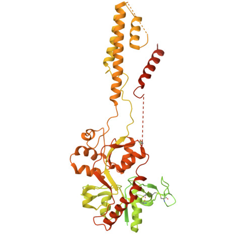
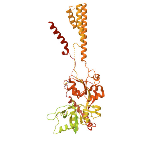
9OEO | pdb_00009oeo

GluN1/GluN3A in complex with glycine, LBD-TMD focused, in desensitized conformation

  • Classification: TRANSPORT PROTEIN
  • Organism(s): Rattus norvegicus
  • Expression System: Homo sapiens
  • Mutation(s): No 

  • Deposited: 2025-04-29 Released: 2026-08-26 
  • Deposition Author(s): Kim, J., Gouaux, E.
  • Funding Organization(s): National Institutes of Health/National Institute of Neurological Disorders and Stroke (NIH/NINDS)

Experimental Data Snapshot

  • Method: ELECTRON MICROSCOPY
  • Resolution: 4.21 Å
  • Aggregation State: PARTICLE 
  • Reconstruction Method: SINGLE PARTICLE 

wwPDB Validation 3D Report Full Report

Validation slider image for 9OEO

This is version 1.1 of the entry. See complete history. 

Literature

Structural and mechanistic insights into gating and allosteric modulation of GluN1-GluN3A NMDA receptors.

Kim, J., Benton, A.J., Lotti, J.S., Rouzbeh, N., Hansen, K.B., Gouaux, E.

(2026) Nat Struct Mol Biol 33: 1299-1310

  • DOI: https://doi.org/10.1038/s41594-026-01866-9
  • Primary Citation Related Structures: 
    9OEH, 9OEM, 9OEN, 9OEO, 9OFI, 9OFM, 9OFY, 9OFZ

  • PubMed Abstract: 

    N-methyl-D-aspartate receptors (NMDARs) mediate excitatory signaling essential for synaptic plasticity and memory. Unlike GluN2-containing NMDARs, GluN3-containing receptors are activated solely by glycine and exhibit profound desensitization and paradoxical potentiation by GluN1-selective antagonists, including CGP-78608 (CGP). Although GluN3A-containing NMDARs regulate synapse pruning and excitotoxicity, and are associated with schizophrenia, autism and stroke, their native stoichiometry and gating mechanism are poorly defined. Here, using single-molecule pull-down analysis, we show that native GluN3A-containing receptors are diheteromeric assemblies. Cryogenic-electron microscopy analysis of GluN1-GluN3A receptors in antagonist-bound, preactive, active and desensitized states, augmented by electrophysiology and pharmacology experiments, show how glycine activates the receptor solely via GluN3A-dependent conformational changes, opening the gate with two-fold symmetry, and induces a roughly four-fold symmetric desensitized state. CGP-bound GluN1 restricts GluN3A rotation, promoting glycine-induced activation by blocking desensitization. These findings illuminate how CGP potentiates GluN1-GluN3A receptor activity, place the receptor gating mechanism on a structural foundation and define the molecular basis for pharmacological modulation.


  • Organizational Affiliation: 
    • Vollum Institute, Oregon Health and Science University, Portland, OR, USA.

Macromolecule Content 

  • Total Structure Weight: 419.4 kDa 
  • Atom Count: 10,884 
  • Modeled Residue Count: 1,563 
  • Deposited Residue Count: 3,726 
  • Unique protein chains: 2

Macromolecules

Find similar proteins by:|  3D Structure
Entity ID: 1
MoleculeChains  Sequence LengthOrganismDetailsImage
Isoform E of Glutamate receptor ionotropic, NMDA 1A,
B [auth C]
870Rattus norvegicusMutation(s): 0 
Gene Names: Grin1, Nmdar1
UniProt
Find proteins for P35439 (Rattus norvegicus)
Explore P35439 
Go to UniProtKB:  P35439
Entity Groups
Sequence Clusters30% Identity50% Identity70% Identity90% Identity95% Identity100% Identity
UniProt GroupP35439
Glycosylation
Glycosylation Sites: 2Go to GlyGen: P35439-1
Sequence Annotations
Expand
Reference Sequence
Find similar proteins by:|  3D Structure
Entity ID: 2
MoleculeChains  Sequence LengthOrganismDetailsImage
Glutamate receptor ionotropic, NMDA 3AC [auth B],
D
993Rattus norvegicusMutation(s): 0 
Gene Names: Grin3a
UniProt
Find proteins for Q9R1M7 (Rattus norvegicus)
Explore Q9R1M7 
Go to UniProtKB:  Q9R1M7
Entity Groups
Sequence Clusters30% Identity50% Identity70% Identity90% Identity95% Identity100% Identity
UniProt GroupQ9R1M7
Glycosylation
Glycosylation Sites: 2Go to GlyGen: Q9R1M7-1
Sequence Annotations
Expand
Reference Sequence

Small Molecules

Ligands 1 Unique
IDChains Name / Formula / InChI Key2D Diagram3D Interactions
NAG
(Subject of Investigation/LOI)

Query on NAG


Download:Ideal Coordinates CCD File
E [auth A]
F [auth A]
G [auth C]
H [auth C]
I [auth B]
E [auth A],
F [auth A],
G [auth C],
H [auth C],
I [auth B],
J [auth B],
K [auth B],
L [auth D],
M [auth D],
N [auth D]
2-acetamido-2-deoxy-beta-D-glucopyranose
C8 H15 N O6
OVRNDRQMDRJTHS-FMDGEEDCSA-N

Experimental Data & Validation

Experimental Data

  • Method: ELECTRON MICROSCOPY
  • Resolution: 4.21 Å
  • Aggregation State: PARTICLE 
  • Reconstruction Method: SINGLE PARTICLE 
EM Software:
TaskSoftware PackageVersion
MODEL REFINEMENTPHENIX1.20.
RECONSTRUCTIONcryoSPARC4.4.0

Structure Validation

View Full Validation Report


Entry History 

& Funding Information

Deposition Data


Funding OrganizationLocationGrant Number
National Institutes of Health/National Institute of Neurological Disorders and Stroke (NIH/NINDS)United States--

Revision History  (Full details and data files)

  • Version 1.0: 2026-08-26
    Type: Initial release
  • Version 1.1: 2026-10-21
    Changes: Data collection, Database references