8VQY

Human GABAA receptor alpha1-beta2-gamma2 subtype in complex with GABA plus methaqualone


Experimental Data Snapshot

  • Method: ELECTRON MICROSCOPY
  • Resolution: 2.82 Å
  • Aggregation State: PARTICLE 
  • Reconstruction Method: SINGLE PARTICLE 

wwPDB Validation   3D Report Full Report


This is version 1.1 of the entry. See complete history


Literature

Structural insights into GABA A receptor potentiation by Quaalude.

Chojnacka, W.Teng, J.Kim, J.J.Jensen, A.A.Hibbs, R.E.

(2024) Nat Commun 15: 5244-5244

  • DOI: https://doi.org/10.1038/s41467-024-49471-y
  • Primary Citation of Related Structures:  
    8VQY, 8VRN

  • PubMed Abstract: 

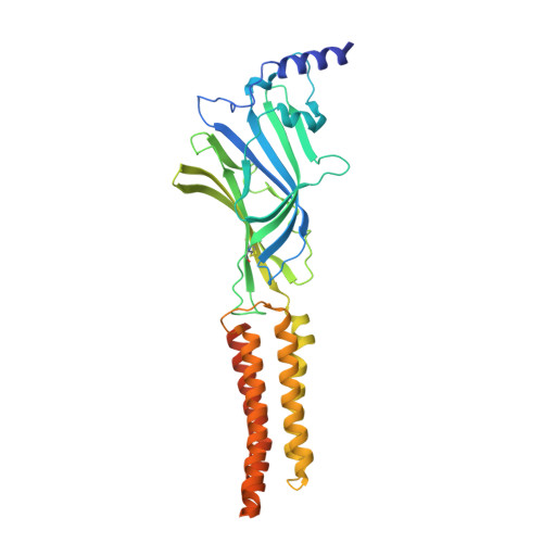
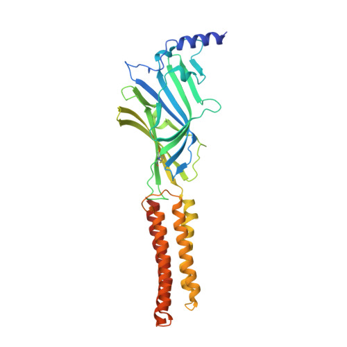
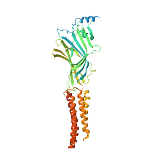
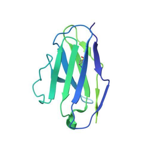
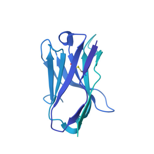
    Methaqualone, a quinazolinone marketed commercially as Quaalude, is a central nervous system depressant that was used clinically as a sedative-hypnotic, then became a notorious recreational drug in the 1960s-80s. Due to its high abuse potential, medical use of methaqualone was eventually prohibited, yet it persists as a globally abused substance. Methaqualone principally targets GABA A receptors, which are the major inhibitory neurotransmitter-gated ion channels in the brain. The restricted status and limited accessibility of methaqualone have contributed to its pharmacology being understudied. Here, we use cryo-EM to localize the GABA A receptor binding sites of methaqualone and its more potent derivative, PPTQ, to the same intersubunit transmembrane sites targeted by the general anesthetics propofol and etomidate. Both methaqualone and PPTQ insert more deeply into subunit interfaces than the previously-characterized modulators. Binding of quinazolinones to this site results in widening of the extracellular half of the ion-conducting pore, following a trend among positive allosteric modulators in destabilizing the hydrophobic activation gate in the pore as a mechanism for receptor potentiation. These insights shed light on the underexplored pharmacology of quinazolinones and further elucidate the molecular mechanisms of allosteric GABA A receptor modulation through transmembrane binding sites.


  • Organizational Affiliation

    Biomedical Sciences Graduate Program, University of California San Diego, La Jolla, CA, USA.


Macromolecules
Find similar proteins by:  (by identity cutoff)  |  3D Structure
Entity ID: 1
MoleculeChains Sequence LengthOrganismDetailsImage
Gamma-aminobutyric acid receptor subunit beta-2
A, C
364Homo sapiensMutation(s): 0 
Gene Names: GABRB2
UniProt & NIH Common Fund Data Resources
Find proteins for P47870 (Homo sapiens)
Explore P47870 
Go to UniProtKB:  P47870
PHAROS:  P47870
GTEx:  ENSG00000145864 
Entity Groups  
Sequence Clusters30% Identity50% Identity70% Identity90% Identity95% Identity100% Identity
UniProt GroupP47870
Glycosylation
Glycosylation Sites: 2Go to GlyGen: P47870-2
Sequence Annotations
Expand
  • Reference Sequence
Find similar proteins by:  (by identity cutoff)  |  3D Structure
Entity ID: 2
MoleculeChains Sequence LengthOrganismDetailsImage
Gamma-aminobutyric acid receptor subunit alpha-1
B, D
358Homo sapiensMutation(s): 0 
Gene Names: GABRA1
UniProt & NIH Common Fund Data Resources
Find proteins for P14867 (Homo sapiens)
Explore P14867 
Go to UniProtKB:  P14867
GTEx:  ENSG00000022355 
Entity Groups  
Sequence Clusters30% Identity50% Identity70% Identity90% Identity95% Identity100% Identity
UniProt GroupP14867
Glycosylation
Glycosylation Sites: 1Go to GlyGen: P14867-1
Sequence Annotations
Expand
  • Reference Sequence
Find similar proteins by:  (by identity cutoff)  |  3D Structure
Entity ID: 3
MoleculeChains Sequence LengthOrganismDetailsImage
Gamma-aminobutyric acid receptor subunit gamma-2417Homo sapiensMutation(s): 0 
Gene Names: GABRG2
UniProt & NIH Common Fund Data Resources
Find proteins for P18507 (Homo sapiens)
Explore P18507 
Go to UniProtKB:  P18507
GTEx:  ENSG00000113327 
Entity Groups  
Sequence Clusters30% Identity50% Identity70% Identity90% Identity95% Identity100% Identity
UniProt GroupP18507
Glycosylation
Glycosylation Sites: 1Go to GlyGen: P18507-2
Sequence Annotations
Expand
  • Reference Sequence
Find similar proteins by:  (by identity cutoff)  |  3D Structure
Entity ID: 4
MoleculeChains Sequence LengthOrganismDetailsImage
Kappa FAB light chainF [auth I],
H [auth L]
213Mus musculusMutation(s): 0 
Gene Names: LC
Entity Groups  
Sequence Clusters30% Identity50% Identity70% Identity90% Identity95% Identity100% Identity
Sequence Annotations
Expand
  • Reference Sequence
Find similar proteins by:  (by identity cutoff)  |  3D Structure
Entity ID: 5
MoleculeChains Sequence LengthOrganismDetailsImage
IGG2B FAB heavy chainG [auth J],
I [auth K]
454Mus musculusMutation(s): 0 
Gene Names: Ighg
Entity Groups  
Sequence Clusters30% Identity50% Identity70% Identity90% Identity95% Identity100% Identity
Sequence Annotations
Expand
  • Reference Sequence
Oligosaccharides

Help

Entity ID: 6
MoleculeChains Length2D Diagram Glycosylation3D Interactions
2-acetamido-2-deoxy-beta-D-glucopyranose-(1-4)-2-acetamido-2-deoxy-beta-D-glucopyranoseJ [auth F],
K [auth G],
O
2N-Glycosylation
Glycosylation Resources
GlyTouCan:  G42666HT
GlyCosmos:  G42666HT
GlyGen:  G42666HT
Entity ID: 7
MoleculeChains Length2D Diagram Glycosylation3D Interactions
alpha-D-mannopyranose-(1-3)-[alpha-D-mannopyranose-(1-6)]beta-D-mannopyranose-(1-4)-2-acetamido-2-deoxy-beta-D-glucopyranose-(1-4)-2-acetamido-2-deoxy-beta-D-glucopyranoseL [auth H],
N,
P
5N-Glycosylation
Glycosylation Resources
GlyTouCan:  G22768VO
GlyCosmos:  G22768VO
GlyGen:  G22768VO
Entity ID: 8
MoleculeChains Length2D Diagram Glycosylation3D Interactions
alpha-D-mannopyranose-(1-2)-alpha-D-mannopyranose-(1-2)-alpha-D-mannopyranose-(1-3)-[alpha-D-mannopyranose-(1-2)-alpha-D-mannopyranose-(1-6)-[alpha-D-mannopyranose-(1-3)]alpha-D-mannopyranose-(1-6)]beta-D-mannopyranose-(1-4)-2-acetamido-2-deoxy-beta-D-glucopyranose-(1-4)-2-acetamido-2-deoxy-beta-D-glucopyranose
M
10N-Glycosylation
Glycosylation Resources
GlyTouCan:  G40702WU
GlyCosmos:  G40702WU
GlyGen:  G40702WU
Small Molecules
Ligands 4 Unique
IDChains Name / Formula / InChI Key2D Diagram3D Interactions
Q3G
Query on Q3G

Download Ideal Coordinates CCD File 
X [auth C],
Y [auth D]
O-[(R)-[(2S)-2-(hexadecanoyloxy)-3-(octadecanoyloxy)propoxy](hydroxy)phosphoryl]-D-serine
C40 H78 N O10 P
WZFUPCSEUKNOBF-PQQNNWGCSA-N
PTY
Query on PTY

Download Ideal Coordinates CCD File 
S [auth A],
T [auth B],
W [auth C],
Z [auth D]
PHOSPHATIDYLETHANOLAMINE
C40 H80 N O8 P
NJGIRBISCGPRPF-KXQOOQHDSA-N
A1ADG (Subject of Investigation/LOI)
Query on A1ADG

Download Ideal Coordinates CCD File 
AA [auth E],
R [auth A],
V [auth C]
2-methyl-3-(2-methylphenyl)quinazolin-4(3H)-one
C16 H14 N2 O
JEYCTXHKTXCGPB-UHFFFAOYSA-N
ABU
Query on ABU

Download Ideal Coordinates CCD File 
Q [auth A],
U [auth C]
GAMMA-AMINO-BUTANOIC ACID
C4 H9 N O2
BTCSSZJGUNDROE-UHFFFAOYSA-N
Experimental Data & Validation

Experimental Data

  • Method: ELECTRON MICROSCOPY
  • Resolution: 2.82 Å
  • Aggregation State: PARTICLE 
  • Reconstruction Method: SINGLE PARTICLE 

Structure Validation

View Full Validation Report


Entry History & Funding Information

Deposition Data


Funding OrganizationLocationGrant Number
National Institutes of Health/National Institute on Drug Abuse (NIH/NIDA)United StatesDA047325

Revision History  (Full details and data files)

  • Version 1.0: 2024-06-26
    Type: Initial release
  • Version 1.1: 2024-07-03
    Changes: Data collection, Database references