8RXG

Crystal structure of alpha-keto C-methyl transferase SgvM bound to SAM


Experimental Data Snapshot

  • Method: X-RAY DIFFRACTION
  • Resolution: 1.81 Å
  • R-Value Free: 0.237 
  • R-Value Work: 0.195 
  • R-Value Observed: 0.197 

Starting Model: experimental
View more details

wwPDB Validation   3D Report Full Report


Ligand Structure Quality Assessment 


This is version 1.0 of the entry. See complete history


Literature

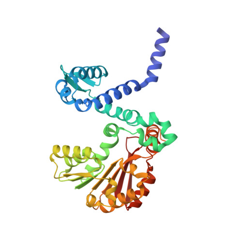
Structures and Protein Engineering of the alpha-Keto Acid C-Methyltransferases SgvM and MrsA for Rational Substrate Transfer.

Sommer-Kamann, C.Breiltgens, J.Zou, Z.Gerhardt, S.Saleem-Batcha, R.Kemper, F.Einsle, O.Andexer, J.N.Muller, M.

(2024) Chembiochem : e202400258-e202400258

  • DOI: https://doi.org/10.1002/cbic.202400258
  • Primary Citation of Related Structures:  
    8R4Z, 8RPR, 8RVC, 8RVS, 8RWM, 8RWW, 8RXF, 8RXG

  • PubMed Abstract: 

    S‑adenosyl-l-methionine-dependent methyltransferases (MTs) are involved in the C-methylation of a variety of natural products. The MTs SgvM from Streptomyces griseoviridis and MrsA from Pseudomonas syringae pv. syringae catalyze the methylation of the β-carbon atom of α-keto acids in the biosynthesis of the antibiotic natural products viridogrisein and 3‑methylarginine, respectively. MrsA shows high substrate selectivity for 5‑guanidino-2-oxovalerate, while other α-keto acids, such as the SgvM substrates 4-methyl-2-oxovalerate, 2-oxovalerate, and phenylpyruvate, are not accepted. Here we report the crystal structures of SgvM and MrsA in the apo form bound with substrate or S‑adenosyl-l-methionine. By investigating key residues for substrate recognition in the active sites of both enzymes and engineering MrsA by site-directed mutagenesis, the substrate range of MrsA was extended to accept α‑keto acid substrates of SgvM with uncharged and lipophilic β‑residues. Our results showcase the transfer of the substrate scope of α-keto acid MTs from different biosynthetic pathways by rational design.


  • Organizational Affiliation

    University of Freiburg, Institute of pharmaceutical science, GERMANY.


Macromolecules
Find similar proteins by:  (by identity cutoff)  |  3D Structure
Entity ID: 1
MoleculeChains Sequence LengthOrganismDetailsImage
Methyltransferase339Streptomyces griseoviridisMutation(s): 0 
UniProt
Find proteins for R9UTR3 (Streptomyces griseoviridis)
Explore R9UTR3 
Go to UniProtKB:  R9UTR3
Entity Groups  
Sequence Clusters30% Identity50% Identity70% Identity90% Identity95% Identity100% Identity
UniProt GroupR9UTR3
Sequence Annotations
Expand
  • Reference Sequence
Small Molecules
Ligands 4 Unique
IDChains Name / Formula / InChI Key2D Diagram3D Interactions
SAM
Query on SAM

Download Ideal Coordinates CCD File 
E [auth A]S-ADENOSYLMETHIONINE
C15 H22 N6 O5 S
MEFKEPWMEQBLKI-FCKMPRQPSA-N
COI (Subject of Investigation/LOI)
Query on COI

Download Ideal Coordinates CCD File 
C [auth A]2-OXO-4-METHYLPENTANOIC ACID
C6 H10 O3
BKAJNAXTPSGJCU-UHFFFAOYSA-N
ZN
Query on ZN

Download Ideal Coordinates CCD File 
B [auth A]ZINC ION
Zn
PTFCDOFLOPIGGS-UHFFFAOYSA-N
CL
Query on CL

Download Ideal Coordinates CCD File 
D [auth A]CHLORIDE ION
Cl
VEXZGXHMUGYJMC-UHFFFAOYSA-M
Experimental Data & Validation

Experimental Data

  • Method: X-RAY DIFFRACTION
  • Resolution: 1.81 Å
  • R-Value Free: 0.237 
  • R-Value Work: 0.195 
  • R-Value Observed: 0.197 
  • Space Group: P 43 21 2
Unit Cell:
Length ( Å )Angle ( ˚ )
a = 67.205α = 90
b = 67.205β = 90
c = 183.006γ = 90
Software Package:
Software NamePurpose
BUSTERrefinement
autoPROCdata reduction
XDSdata reduction
autoPROCdata scaling
Aimlessdata scaling
PHASERphasing

Structure Validation

View Full Validation Report


Ligand Structure Quality Assessment 


Entry History & Funding Information

Deposition Data


Funding OrganizationLocationGrant Number
Not funded--

Revision History  (Full details and data files)

  • Version 1.0: 2024-07-24
    Type: Initial release