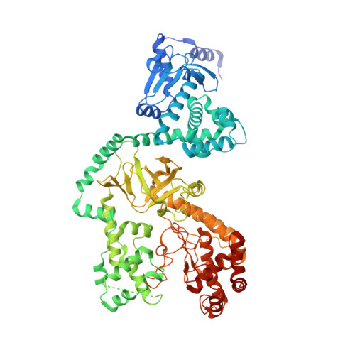
6WJJ

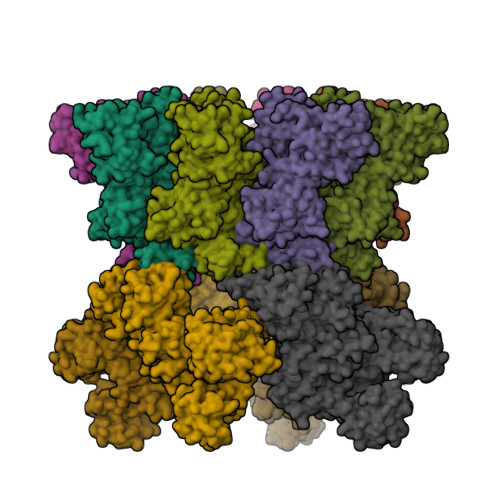
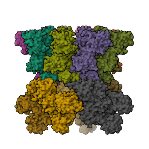
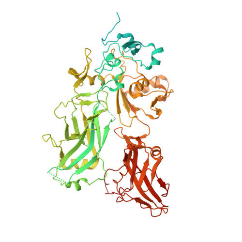
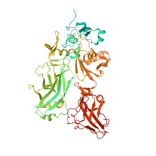
Anthrax octamer prechannel bound to full-length lethal factor


Experimental Data Snapshot

  • Method: ELECTRON MICROSCOPY
  • Resolution: 3.80 Å
  • Aggregation State: PARTICLE 
  • Reconstruction Method: SINGLE PARTICLE 

wwPDB Validation   3D Report Full Report


This is version 1.1 of the entry. See complete history


Literature

Atomic Structures of Anthrax Prechannel Bound with Full-Length Lethal and Edema Factors.

Zhou, K.Liu, S.Hardenbrook, N.J.Cui, Y.Krantz, B.A.Zhou, Z.H.

(2020) Structure 28: 879-887.e3

  • DOI: https://doi.org/10.1016/j.str.2020.05.009
  • Primary Citation of Related Structures:  
    6VRA, 6WJJ

  • PubMed Abstract: 

    Pathogenesis of anthrax disease involves two cytotoxic enzymes-edema factor (EF) and lethal factor (LF)-which are individually recruited by the protective antigen heptamer (PA 7 ) or octamer (PA 8 ) prechannel and subsequently translocated across channels formed on the endosomal membrane upon exposure to low pH. Here, we report the atomic structures of PA 8 prechannel-bound full-length EF and LF. In this pretranslocation state, the N-terminal segment of both factors refolds into an α helix engaged in the α clamp of the prechannel. Recruitment to the PA prechannel exposes an originally buried β strand of both toxins and enables domain organization of EF. Many interactions occur on domain interfaces in both PA prechannel-bound EF and LF, leading to toxin compaction prior to translocation. Our results provide key insights into the molecular mechanisms of translocation-coupled protein unfolding and translocation.


  • Organizational Affiliation

    California NanoSystems Institute, University of California, Los Angeles, CA 90095, USA.


Macromolecules
Find similar proteins by:  (by identity cutoff)  |  3D Structure
Entity ID: 1
MoleculeChains Sequence LengthOrganismDetailsImage
Protective antigenA,
D [auth C],
G [auth D],
J [auth E]
735Bacillus anthracisMutation(s): 2 
Gene Names: pagApagpXO1-110BXA0164GBAA_pXO1_0164
Membrane Entity: Yes 
UniProt
Find proteins for P13423 (Bacillus anthracis)
Explore P13423 
Go to UniProtKB:  P13423
Entity Groups  
Sequence Clusters30% Identity50% Identity70% Identity90% Identity95% Identity100% Identity
UniProt GroupP13423
Sequence Annotations
Expand
  • Reference Sequence
Find similar proteins by:  (by identity cutoff)  |  3D Structure
Entity ID: 2
MoleculeChains Sequence LengthOrganismDetailsImage
Protective antigenB,
E [auth F],
H [auth G],
K [auth H]
735Bacillus anthracisMutation(s): 1 
Gene Names: pagApagpXO1-110BXA0164GBAA_pXO1_0164
Membrane Entity: Yes 
UniProt
Find proteins for P13423 (Bacillus anthracis)
Explore P13423 
Go to UniProtKB:  P13423
Entity Groups  
Sequence Clusters30% Identity50% Identity70% Identity90% Identity95% Identity100% Identity
UniProt GroupP13423
Sequence Annotations
Expand
  • Reference Sequence
Find similar proteins by:  (by identity cutoff)  |  3D Structure
Entity ID: 3
MoleculeChains Sequence LengthOrganismDetailsImage
Lethal factorC [auth L],
F [auth I],
I [auth J],
L [auth K]
776Bacillus anthracisMutation(s): 0 
Gene Names: lef
EC: 3.4.24.83
Membrane Entity: Yes 
UniProt
Find proteins for P15917 (Bacillus anthracis)
Explore P15917 
Go to UniProtKB:  P15917
Entity Groups  
Sequence Clusters30% Identity50% Identity70% Identity90% Identity95% Identity100% Identity
UniProt GroupP15917
Sequence Annotations
Expand
  • Reference Sequence
Small Molecules
Ligands 3 Unique
IDChains Name / Formula / InChI Key2D Diagram3D Interactions
SO4
Query on SO4

Download Ideal Coordinates CCD File 
CA [auth J],
IA [auth K],
Q [auth L],
W [auth I]
SULFATE ION
O4 S
QAOWNCQODCNURD-UHFFFAOYSA-L
ZN
Query on ZN

Download Ideal Coordinates CCD File 
DA [auth J],
JA [auth K],
R [auth L],
X [auth I]
ZINC ION
Zn
PTFCDOFLOPIGGS-UHFFFAOYSA-N
CA
Query on CA

Download Ideal Coordinates CCD File 
AA [auth G]
BA [auth G]
EA [auth E]
FA [auth E]
GA [auth H]
AA [auth G],
BA [auth G],
EA [auth E],
FA [auth E],
GA [auth H],
HA [auth H],
M [auth A],
N [auth A],
O [auth B],
P [auth B],
S [auth C],
T [auth C],
U [auth F],
V [auth F],
Y [auth D],
Z [auth D]
CALCIUM ION
Ca
BHPQYMZQTOCNFJ-UHFFFAOYSA-N
Experimental Data & Validation

Experimental Data

  • Method: ELECTRON MICROSCOPY
  • Resolution: 3.80 Å
  • Aggregation State: PARTICLE 
  • Reconstruction Method: SINGLE PARTICLE 
EM Software:
TaskSoftware PackageVersion
MODEL REFINEMENTPHENIX
MODEL REFINEMENTCoot
RECONSTRUCTIONRELION3.0

Structure Validation

View Full Validation Report


Entry History & Funding Information

Deposition Data


Funding OrganizationLocationGrant Number
National Science Foundation (NSF, United States)United StatesDMR-1548924
National Institutes of Health/National Library of Medicine (NIH/NLM)United StatesR01GM071940/AI094386/DE025567
National Institutes of Health/National Library of Medicine (NIH/NLM)United StatesR21AI124020

Revision History  (Full details and data files)

  • Version 1.0: 2020-12-16
    Type: Initial release
  • Version 1.1: 2024-03-06
    Changes: Data collection, Database references