6VRA | pdb_00006vra

Anthrax octamer prechannel bound to full-length edema factor


Experimental Data Snapshot

  • Method: ELECTRON MICROSCOPY
  • Resolution: 3.30 Å
  • Aggregation State: PARTICLE 
  • Reconstruction Method: SINGLE PARTICLE 

wwPDB Validation 3D Report Full Report

Validation slider image for 6VRA

This is version 1.1 of the entry. See complete history

Literature

Atomic Structures of Anthrax Prechannel Bound with Full-Length Lethal and Edema Factors.

Zhou, K.Liu, S.Hardenbrook, N.J.Cui, Y.Krantz, B.A.Zhou, Z.H.

(2020) Structure 28: 879-887.e3

  • DOI: https://doi.org/10.1016/j.str.2020.05.009
  • Primary Citation Related Structures: 
    6VRA, 6WJJ

  • PubMed Abstract: 

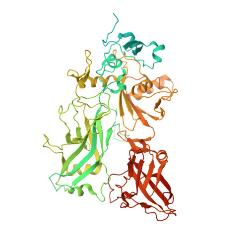
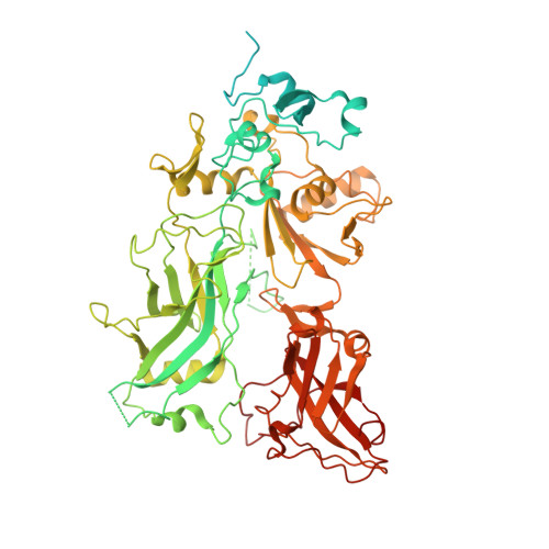
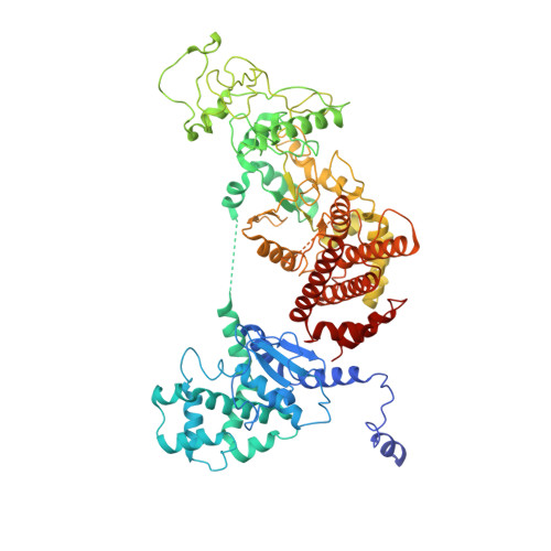
    Pathogenesis of anthrax disease involves two cytotoxic enzymes-edema factor (EF) and lethal factor (LF)-which are individually recruited by the protective antigen heptamer (PA 7 ) or octamer (PA 8 ) prechannel and subsequently translocated across channels formed on the endosomal membrane upon exposure to low pH. Here, we report the atomic structures of PA 8 prechannel-bound full-length EF and LF. In this pretranslocation state, the N-terminal segment of both factors refolds into an α helix engaged in the α clamp of the prechannel. Recruitment to the PA prechannel exposes an originally buried β strand of both toxins and enables domain organization of EF. Many interactions occur on domain interfaces in both PA prechannel-bound EF and LF, leading to toxin compaction prior to translocation. Our results provide key insights into the molecular mechanisms of translocation-coupled protein unfolding and translocation.


  • Organizational Affiliation
    • California NanoSystems Institute, University of California, Los Angeles, CA 90095, USA.

Macromolecule Content 

  • Total Structure Weight: 1,017.07 kDa 
  • Atom Count: 57,744 
  • Modeled Residue Count: 7,208 
  • Deposited Residue Count: 8,948 
  • Unique protein chains: 3

Macromolecules

Find similar proteins by:|  3D Structure
Entity ID: 1
MoleculeChains  Sequence LengthOrganismDetailsImage
Protective antigen
A, C, E, G
735Bacillus anthracisMutation(s): 2 
Gene Names: pagApagpXO1-110BXA0164GBAA_pXO1_0164
Membrane Entity: Yes 
UniProt
Find proteins for P13423 (Bacillus anthracis)
Explore P13423 
Go to UniProtKB:  P13423
Entity Groups
Sequence Clusters30% Identity50% Identity70% Identity90% Identity95% Identity100% Identity
UniProt GroupP13423
Sequence Annotations
Expand
Reference Sequence
Find similar proteins by:|  3D Structure
Entity ID: 2
MoleculeChains  Sequence LengthOrganismDetailsImage
Protective antigen
B, D, F, H
735Bacillus anthracisMutation(s): 1 
Gene Names: pagApagpXO1-110BXA0164GBAA_pXO1_0164
Membrane Entity: Yes 
UniProt
Find proteins for P13423 (Bacillus anthracis)
Explore P13423 
Go to UniProtKB:  P13423
Entity Groups
Sequence Clusters30% Identity50% Identity70% Identity90% Identity95% Identity100% Identity
UniProt GroupP13423
Sequence Annotations
Expand
Reference Sequence
Find similar proteins by:|  3D Structure
Entity ID: 3
MoleculeChains  Sequence LengthOrganismDetailsImage
Calmodulin-sensitive adenylate cyclase
I, J, K, L
767Bacillus anthracisMutation(s): 0 
Gene Names: cyapXO1-122BXA0141GBAA_pXO1_0142
EC: 4.6.1.1
Membrane Entity: Yes 
UniProt
Find proteins for P40136 (Bacillus anthracis)
Explore P40136 
Go to UniProtKB:  P40136
Entity Groups
Sequence Clusters30% Identity50% Identity70% Identity90% Identity95% Identity100% Identity
UniProt GroupP40136
Sequence Annotations
Expand
Reference Sequence

Small Molecules

Ligands 1 Unique
IDChains Name / Formula / InChI Key2D Diagram3D Interactions
CA

Query on CA


Download:Ideal Coordinates CCD File
AA [auth H]
BA [auth H]
M [auth A]
N [auth A]
O [auth B]
AA [auth H],
BA [auth H],
M [auth A],
N [auth A],
O [auth B],
P [auth B],
Q [auth C],
R [auth C],
S [auth D],
T [auth D],
U [auth E],
V [auth E],
W [auth F],
X [auth F],
Y [auth G],
Z [auth G]
CALCIUM ION
Ca
BHPQYMZQTOCNFJ-UHFFFAOYSA-N

Experimental Data & Validation

Experimental Data

  • Method: ELECTRON MICROSCOPY
  • Resolution: 3.30 Å
  • Aggregation State: PARTICLE 
  • Reconstruction Method: SINGLE PARTICLE 
EM Software:
TaskSoftware PackageVersion
MODEL REFINEMENTPHENIX
MODEL REFINEMENTCoot
RECONSTRUCTIONRELION3.0

Structure Validation

View Full Validation Report


Entry History 

& Funding Information

Deposition Data


Funding OrganizationLocationGrant Number
National Science Foundation (NSF, United States)United StatesDMR-1548924
National Institutes of Health/National Library of Medicine (NIH/NLM)United StatesR01GM071940/AI094386/DE025567
National Institutes of Health/National Library of Medicine (NIH/NLM)United StatesR21AI124020

Revision History  (Full details and data files)

  • Version 1.0: 2020-12-16
    Type: Initial release
  • Version 1.1: 2024-03-06
    Changes: Data collection, Database references