6BI4 | pdb_00006bi4

2.9 Angstrom Resolution Crystal Structure of dTDP-Glucose 4,6-dehydratase (rfbB) from Bacillus anthracis str. Ames in Complex with NAD.


Experimental Data Snapshot

  • Method: X-RAY DIFFRACTION
  • Resolution: 2.91 Å
  • R-Value Free: 
    0.204 (Depositor), 0.213 (DCC) 
  • R-Value Work: 
    0.167 (Depositor), 0.175 (DCC) 
  • R-Value Observed: 
    0.169 (Depositor) 

Starting Model: experimental
View more details

wwPDB Validation 3D Report Full Report

Validation slider image for 6BI4

Ligand Structure Quality Assessment 


This is version 2.4 of the entry. See complete history

Literature

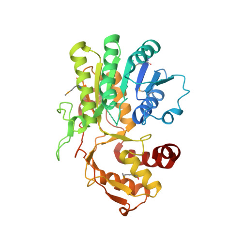
Structure of the Bacillus anthracis dTDP-l-rhamnose biosynthetic pathway enzyme: dTDP-alpha-d-glucose 4,6-dehydratase, RfbB.

Gokey, T.Halavaty, A.S.Minasov, G.Anderson, W.F.Kuhn, M.L.

(2018) J Struct Biol 202: 175-181

  • DOI: https://doi.org/10.1016/j.jsb.2018.01.006
  • Primary Citation Related Structures: 
    6BI4

  • PubMed Abstract: 

    Many bacteria require l-rhamnose as a key cell wall component. This sugar is transferred to the cell wall using an activated donor dTDP-l-rhamnose, which is produced by the dTDP-l-rhamnose biosynthetic pathway. We determined the crystal structure of the second enzyme of this pathway dTDP-α-d-glucose 4,6-dehydratase (RfbB) from Bacillus anthracis. Interestingly, RfbB only crystallized in the presence of the third enzyme of the pathway RfbC; however, RfbC was not present in the crystal. Our work represents the first complete structural characterization of the four proteins of this pathway in a single Gram-positive bacterium.


  • Organizational Affiliation
    • Department of Chemistry and Biochemistry, San Francisco State University, USA.

Macromolecule Content 

  • Total Structure Weight: 164.52 kDa 
  • Atom Count: 10,407 
  • Modeled Residue Count: 1,242 
  • Deposited Residue Count: 1,384 
  • Unique protein chains: 1

Macromolecules

Find similar proteins by:|  3D Structure
Entity ID: 1
MoleculeChains  Sequence LengthOrganismDetailsImage
dTDP-glucose 4,6-dehydratase
A, B, C, D
346Bacillus anthracis str. AmesMutation(s): 0 
Gene Names: rfbB
EC: 4.2.1.46
UniProt
Find proteins for A0A6L7HMN5 (Bacillus anthracis)
Explore A0A6L7HMN5 
Go to UniProtKB:  A0A6L7HMN5
Entity Groups
Sequence Clusters30% Identity50% Identity70% Identity90% Identity95% Identity100% Identity
UniProt GroupA0A6L7HMN5
Sequence Annotations
Expand
Reference Sequence

Oligosaccharides

Help  
Entity ID: 2
MoleculeChains Length2D Diagram GlycosylationD Interactions
beta-D-fructofuranose-(2-1)-alpha-D-glucopyranose
E, F
2N/A
Glycosylation Resources
GlyTouCan: G05551OP
GlyCosmos: G05551OP

Small Molecules

Ligands 3 Unique
IDChains Name / Formula / InChI Key2D Diagram3D Interactions
NAD

Query on NAD


Download:Ideal Coordinates CCD File
H [auth A],
M [auth B],
R [auth C],
V [auth D]
NICOTINAMIDE-ADENINE-DINUCLEOTIDE
C21 H27 N7 O14 P2
BAWFJGJZGIEFAR-NNYOXOHSSA-N
SO4

Query on SO4


Download:Ideal Coordinates CCD File
I [auth A]
J [auth A]
K [auth A]
N [auth B]
O [auth B]
I [auth A],
J [auth A],
K [auth A],
N [auth B],
O [auth B],
P [auth B],
Q [auth B],
S [auth C],
T [auth C],
U [auth C],
W [auth D],
X [auth D],
Y [auth D],
Z [auth D]
SULFATE ION
O4 S
QAOWNCQODCNURD-UHFFFAOYSA-L
NI

Query on NI


Download:Ideal Coordinates CCD File
G [auth A],
L [auth B]
NICKEL (II) ION
Ni
VEQPNABPJHWNSG-UHFFFAOYSA-N
Modified Residues  1 Unique
IDChains TypeFormula2D DiagramParent
MSE
Query on MSE
A, B, C, D
L-PEPTIDE LINKINGC5 H11 N O2 SeMET

Biologically Interesting Molecules (External Reference) 

1 Unique

Experimental Data & Validation

Experimental Data

  • Method: X-RAY DIFFRACTION
  • Resolution: 2.91 Å
  • R-Value Free:  0.204 (Depositor), 0.213 (DCC) 
  • R-Value Work:  0.167 (Depositor), 0.175 (DCC) 
  • R-Value Observed: 0.169 (Depositor) 
Space Group: I 4 2 2
Unit Cell:
Length ( Å )Angle ( ˚ )
a = 165.557α = 90
b = 165.557β = 90
c = 292.121γ = 90
Software Package:
Software NamePurpose
REFMACrefinement
HKL-2000data reduction
HKL-2000data scaling
PHASERphasing

Structure Validation

View Full Validation Report


Ligand Structure Quality Assessment 


Entry History 

Deposition Data

Revision History  (Full details and data files)

  • Version 1.0: 2017-11-08
    Type: Initial release
  • Version 2.0: 2020-07-29
    Type: Remediation
    Reason: Carbohydrate remediation
    Changes: Atomic model, Data collection, Derived calculations, Non-polymer description, Structure summary
  • Version 2.1: 2021-01-27
    Changes: Database references, Structure summary
  • Version 2.2: 2023-10-04
    Changes: Data collection, Database references, Refinement description
  • Version 2.3: 2023-11-15
    Changes: Data collection
  • Version 2.4: 2024-10-16
    Changes: Structure summary