4AAB | pdb_00004aab

Crystal structure of the mutant D75N I-CreI in complex with its wild- type target (The four central bases, 2NN region, are composed by GTAC from 5' to 3')


Experimental Data Snapshot

  • Method: X-RAY DIFFRACTION
  • Resolution: 2.50 Å
  • R-Value Free: 
    0.225 (Depositor), 0.213 (DCC) 
  • R-Value Work: 
    0.170 (Depositor), 0.161 (DCC) 
  • R-Value Observed: 
    0.173 (Depositor) 

Starting Model: experimental
View more details

wwPDB Validation 3D Report Full Report

Validation slider image for 4AAB

This is version 1.2 of the entry. See complete history

Literature

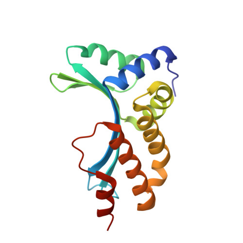
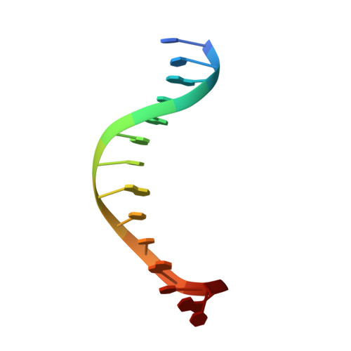
Non-Specific Protein-DNA Interactions Control I-Crei Target Binding and Cleavage.

Molina, R.Redondo, P.Stella, S.Marenchino, M.D'Abramo, M.Gervasio, F.L.Charles Epinat, J.Valton, J.Grizot, S.Duchateau, P.Prieto, J.Montoya, G.

(2012) Nucleic Acids Res 40: 6936-6945

  • DOI: https://doi.org/10.1093/nar/gks320
  • Primary Citation Related Structures: 
    4AAB, 4AAD, 4AAE, 4AAF, 4AAG

  • PubMed Abstract: 

    Homing endonucleases represent protein scaffolds that provide powerful tools for genome manipulation, as these enzymes possess a very low frequency of DNA cleavage in eukaryotic genomes due to their high specificity. The basis of protein-DNA recognition must be understood to generate tailored enzymes that target the DNA at sites of interest. Protein-DNA interaction engineering of homing endonucleases has demonstrated the potential of these approaches to create new specific instruments to target genes for inactivation or repair. Protein-DNA interface studies have been focused mostly on specific contacts between amino acid side chains and bases to redesign the binding interface. However, it has been shown that 4 bp in the central DNA sequence of the 22-bp substrate of a homing endonuclease (I-CreI), which do not show specific protein-DNA interactions, is not devoid of content information. Here, we analyze the mechanism of target discrimination in this substrate region by the I-CreI protein, determining how it can occur independently of the specific protein-DNA interactions. Our data suggest the important role of indirect readout in this substrate region, opening the possibility for a fully rational search of new target sequences, thus improving the development of redesigned enzymes for therapeutic and biotechnological applications.


  • Organizational Affiliation
    • Structural Biology and Biocomputing Programme, Spanish National Cancer Research Centre (CNIO), Macromolecular Crystallography Group, Madrid, Spain.

Macromolecule Content 

  • Total Structure Weight: 50.15 kDa 
  • Atom Count: 3,603 
  • Modeled Residue Count: 352 
  • Deposited Residue Count: 352 
  • Unique protein chains: 1
  • Unique nucleic acid chains: 2

Macromolecules


Find similar proteins by:|  3D Structure
Entity ID: 1
MoleculeChains  Sequence LengthOrganismDetailsImage
DNA ENDONUCLEASE I-CREI
A, B
152Chlamydomonas reinhardtiiMutation(s): 1 
EC: 3.1
UniProt
Find proteins for P05725 (Chlamydomonas reinhardtii)
Explore P05725 
Go to UniProtKB:  P05725
Entity Groups
Sequence Clusters30% Identity50% Identity70% Identity90% Identity95% Identity100% Identity
UniProt GroupP05725
Sequence Annotations
Expand
Reference Sequence
Find similar nucleic acids by:  Sequence
Entity ID: 2
MoleculeChains LengthOrganismImage
14MER DNA 5'-D(*TP*CP*AP*AP*AP*AP*CP*GP*TP*CP*GP*TP*AP*CP)-3'C [auth D],
E [auth F]
14synthetic construct
Sequence Annotations
Expand
Reference Sequence
Find similar nucleic acids by:  Sequence
Entity ID: 3
MoleculeChains LengthOrganismImage
10MER DNA 5'-D(*GP*AP*CP*GP*TP*TP*TP*TP*GP*AP)-3'D [auth E],
F [auth G]
10synthetic construct
Sequence Annotations
Expand
Reference Sequence

Small Molecules

Ligands 3 Unique
IDChains Name / Formula / InChI Key2D Diagram3D Interactions
PGO

Query on PGO


Download:Ideal Coordinates CCD File
G [auth B],
H [auth B],
K [auth D],
L [auth E],
M [auth E]
S-1,2-PROPANEDIOL
C3 H8 O2
DNIAPMSPPWPWGF-VKHMYHEASA-N
MG

Query on MG


Download:Ideal Coordinates CCD File
I [auth B],
J [auth B],
N [auth E]
MAGNESIUM ION
Mg
JLVVSXFLKOJNIY-UHFFFAOYSA-N
NA

Query on NA


Download:Ideal Coordinates CCD File
O [auth G]SODIUM ION
Na
FKNQFGJONOIPTF-UHFFFAOYSA-N

Experimental Data & Validation

Experimental Data

  • Method: X-RAY DIFFRACTION
  • Resolution: 2.50 Å
  • R-Value Free:  0.225 (Depositor), 0.213 (DCC) 
  • R-Value Work:  0.170 (Depositor), 0.161 (DCC) 
  • R-Value Observed: 0.173 (Depositor) 
Space Group: P 21 21 2
Unit Cell:
Length ( Å )Angle ( ˚ )
a = 171.97α = 90
b = 71.1β = 90
c = 46.92γ = 90
Software Package:
Software NamePurpose
PHENIXrefinement
XDSdata reduction
SCALAdata scaling
PHASERphasing

Structure Validation

View Full Validation Report


Entry History 

Revision History  (Full details and data files)

  • Version 1.0: 2012-05-02
    Type: Initial release
  • Version 1.1: 2012-08-22
    Changes: Database references
  • Version 1.2: 2023-12-20
    Changes: Data collection, Database references, Derived calculations, Other, Refinement description