3GEW | pdb_00003gew

FaeE-FaeG chaperone-major pilin complex of F4 ad fimbriae


Experimental Data Snapshot

  • Method: X-RAY DIFFRACTION
  • Resolution: 2.00 Å
  • R-Value Free: 
    0.261 (Depositor), 0.254 (DCC) 
  • R-Value Work: 
    0.223 (Depositor) 
  • R-Value Observed: 
    0.225 (Depositor) 

Starting Models: experimental
View more details

wwPDB Validation 3D Report Full Report

Validation slider image for 3GEW

This is version 1.4 of the entry. See complete history

Literature

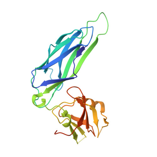
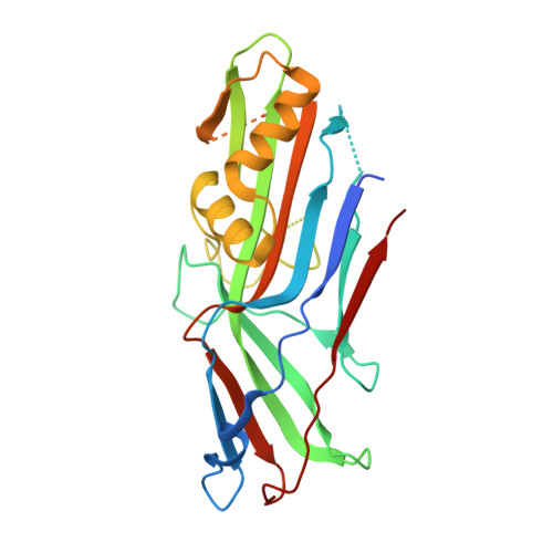
Structural and thermodynamic characterization of pre- and postpolymerization states in the F4 fimbrial subunit FaeG

Van Molle, I.Moonens, K.Garcia-Pino, A.Buts, L.De Kerpel, M.Wyns, L.Bouckaert, J.De Greve, H.

(2009) J Mol Biology 394: 957-967

  • DOI: https://doi.org/10.1016/j.jmb.2009.09.059
  • Primary Citation Related Structures: 
    3GEA, 3GEW, 3GFU, 3GGH, 3HLR

  • PubMed Abstract: 

    Enterotoxigenic Escherichia coli expressing F4 fimbriae are the major cause of porcine colibacillosis and are responsible for significant death and morbidity in neonatal and postweaned piglets. Via the chaperone-usher pathway, F4 fimbriae are assembled into thin, flexible polymers mainly composed of the single-domain adhesin FaeG. The F4 fimbrial system has been labeled eccentric because the F4 pilins show some features distinct from the features of pilins of other chaperone-usher-assembled structures. In particular, FaeG is much larger than other pilins (27 versus approximately 17 kDa), grafting an additional carbohydrate binding domain on the common immunoglobulin-like core. Structural data of FaeG during different stages of the F4 fimbrial biogenesis process, combined with differential scanning calorimetry measurements, confirm the general principles of the donor strand complementation/exchange mechanisms taking place during pilus biogenesis via the chaperone-usher pathway.


  • Organizational Affiliation
    • Structural Biology Brussels, Vrije Universiteit Brussel, Pleinlaan 2, 1050 Brussels, Belgium.

Macromolecule Content 

  • Total Structure Weight: 104.31 kDa 
  • Atom Count: 6,440 
  • Modeled Residue Count: 839 
  • Deposited Residue Count: 954 
  • Unique protein chains: 2

Macromolecules

Find similar proteins by:|  3D Structure
Entity ID: 1
MoleculeChains  Sequence LengthOrganismDetailsImage
Chaperone protein faeEA [auth B],
B [auth C]
224Escherichia coliMutation(s): 0 
Gene Names: faeE
UniProt
Find proteins for P25401 (Escherichia coli)
Explore P25401 
Go to UniProtKB:  P25401
Entity Groups
Sequence Clusters30% Identity50% Identity70% Identity90% Identity95% Identity100% Identity
UniProt GroupP25401
Sequence Annotations
Expand
Reference Sequence
Find similar proteins by:|  3D Structure
Entity ID: 2
MoleculeChains  Sequence LengthOrganismDetailsImage
K88 fimbrial protein ADC [auth D],
D [auth A]
253Escherichia coliMutation(s): 0 
Gene Names: faeG
UniProt
Find proteins for P14191 (Escherichia coli)
Explore P14191 
Go to UniProtKB:  P14191
Entity Groups
Sequence Clusters30% Identity50% Identity70% Identity90% Identity95% Identity100% Identity
UniProt GroupP14191
Sequence Annotations
Expand
Reference Sequence

Small Molecules

Ligands 2 Unique
IDChains Name / Formula / InChI Key2D Diagram3D Interactions
SO4

Query on SO4


Download:Ideal Coordinates CCD File
E [auth B]
F [auth B]
G [auth B]
H [auth B]
I [auth B]
E [auth B],
F [auth B],
G [auth B],
H [auth B],
I [auth B],
J [auth C],
K [auth C],
L [auth C],
M [auth C],
N [auth C],
O [auth D],
Q [auth A]
SULFATE ION
O4 S
QAOWNCQODCNURD-UHFFFAOYSA-L
GOL

Query on GOL


Download:Ideal Coordinates CCD File
P [auth D],
R [auth A]
GLYCEROL
C3 H8 O3
PEDCQBHIVMGVHV-UHFFFAOYSA-N

Experimental Data & Validation

Experimental Data

  • Method: X-RAY DIFFRACTION
  • Resolution: 2.00 Å
  • R-Value Free:  0.261 (Depositor), 0.254 (DCC) 
  • R-Value Work:  0.223 (Depositor) 
  • R-Value Observed: 0.225 (Depositor) 
Space Group: P 1 21 1
Unit Cell:
Length ( Å )Angle ( ˚ )
a = 74.576α = 90
b = 89.757β = 113.94
c = 85.826γ = 90
Software Package:
Software NamePurpose
PHASERphasing
PHENIXrefinement
PDB_EXTRACTdata extraction
DENZOdata reduction
SCALEPACKdata scaling

Structure Validation

View Full Validation Report


Entry History 

Deposition Data

Revision History  (Full details and data files)

  • Version 1.0: 2009-10-20
    Type: Initial release
  • Version 1.1: 2011-07-13
    Changes: Non-polymer description, Version format compliance
  • Version 1.2: 2014-01-29
    Changes: Database references
  • Version 1.3: 2017-08-09
    Changes: Refinement description, Source and taxonomy
  • Version 1.4: 2023-11-01
    Changes: Data collection, Database references, Derived calculations, Refinement description