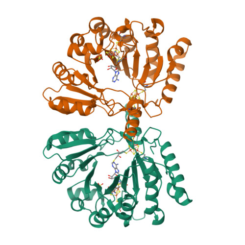
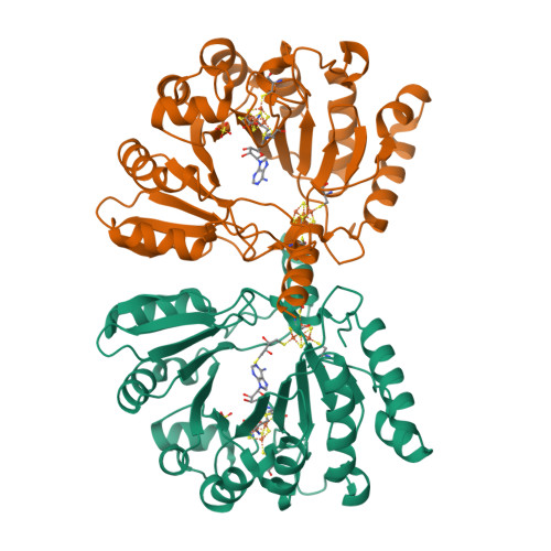
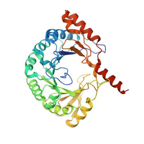
1TV8

Structure of MoaA in complex with S-adenosylmethionine


Experimental Data Snapshot

  • Method: X-RAY DIFFRACTION
  • Resolution: 2.20 Å
  • R-Value Free: 0.241 
  • R-Value Work: 0.213 
  • R-Value Observed: 0.214 

wwPDB Validation   3D Report Full Report


Ligand Structure Quality Assessment 


This is version 1.3 of the entry. See complete history


Literature

Crystal structure of the S-adenosylmethionine-dependent enzyme MoaA and its implications for molybdenum cofactor deficiency in humans.

Hanzelmann, P.Schindelin, H.

(2004) Proc Natl Acad Sci U S A 101: 12870-12875

  • DOI: https://doi.org/10.1073/pnas.0404624101
  • Primary Citation of Related Structures:  
    1TV7, 1TV8

  • PubMed Abstract: 

    The MoaA and MoaC proteins catalyze the first step during molybdenum cofactor biosynthesis, the conversion of a guanosine derivative to precursor Z. MoaA belongs to the S-adenosylmethionine (SAM)-dependent radical enzyme superfamily, members of which catalyze the formation of protein and/or substrate radicals by reductive cleavage of SAM by a [4Fe-4S] cluster. A defined in vitro system is described, which generates precursor Z and led to the identification of 5'-GTP as the substrate. The structures of MoaA in the apo-state (2.8 angstroms) and in complex with SAM (2.2 angstroms) provide valuable insights into its mechanism and help to define the defects caused by mutations in the human ortholog of MoaA that lead to molybdenum cofactor deficiency, a usually fatal disease accompanied by severe neurological symptoms. The central core of each subunit of the MoaA dimer is an incomplete triosephosphate isomerase barrel formed by the N-terminal part of the protein, which contains the [4Fe-4S] cluster typical for SAM-dependent radical enzymes. SAM is the fourth ligand to the cluster and binds to its unique Fe as an N/O chelate. The lateral opening of the incomplete triosephosphate isomerase barrel is covered by the C-terminal part of the protein containing an additional [4Fe-4S] cluster, which is unique to MoaA proteins. Both FeS clusters are separated by approximately 17 angstroms, with a large active site pocket between. The noncysteinyl-ligated unique Fe site of the C-terminal [4Fe-4S] cluster is proposed to be involved in the binding and activation of 5'-GTP.


  • Organizational Affiliation

    Department of Biochemistry, Center for Structural Biology, State University of New York, Stony Brook, NY 11794-5115, USA.


Macromolecules
Find similar proteins by:  (by identity cutoff)  |  3D Structure
Entity ID: 1
MoleculeChains Sequence LengthOrganismDetailsImage
Molybdenum cofactor biosynthesis protein A
A, B
340Staphylococcus aureusMutation(s): 0 
Gene Names: moaA
EC: 4.1.99.22
UniProt
Find proteins for P65388 (Staphylococcus aureus (strain N315))
Explore P65388 
Go to UniProtKB:  P65388
Entity Groups  
Sequence Clusters30% Identity50% Identity70% Identity90% Identity95% Identity100% Identity
UniProt GroupP65388
Sequence Annotations
Expand
  • Reference Sequence
Small Molecules
Ligands 4 Unique
IDChains Name / Formula / InChI Key2D Diagram3D Interactions
SAM
Query on SAM

Download Ideal Coordinates CCD File 
D [auth A],
I [auth B]
S-ADENOSYLMETHIONINE
C15 H22 N6 O5 S
MEFKEPWMEQBLKI-FCKMPRQPSA-N
SF4
Query on SF4

Download Ideal Coordinates CCD File 
E [auth A],
F [auth A],
J [auth B],
K [auth B]
IRON/SULFUR CLUSTER
Fe4 S4
LJBDFODJNLIPKO-UHFFFAOYSA-N
DTU
Query on DTU

Download Ideal Coordinates CCD File 
G [auth A](2R,3S)-1,4-DIMERCAPTOBUTANE-2,3-DIOL
C4 H10 O2 S2
VHJLVAABSRFDPM-ZXZARUISSA-N
SO4
Query on SO4

Download Ideal Coordinates CCD File 
C [auth A],
H [auth B]
SULFATE ION
O4 S
QAOWNCQODCNURD-UHFFFAOYSA-L
Experimental Data & Validation

Experimental Data

  • Method: X-RAY DIFFRACTION
  • Resolution: 2.20 Å
  • R-Value Free: 0.241 
  • R-Value Work: 0.213 
  • R-Value Observed: 0.214 
  • Space Group: P 21 21 21
Unit Cell:
Length ( Å )Angle ( ˚ )
a = 47.982α = 90
b = 103.305β = 90
c = 189.925γ = 90
Software Package:
Software NamePurpose
DENZOdata reduction
SCALEPACKdata scaling
FFTmodel building
REFMACrefinement
FFTphasing

Structure Validation

View Full Validation Report


Ligand Structure Quality Assessment 


Entry History 

Deposition Data

Revision History  (Full details and data files)

  • Version 1.0: 2004-08-31
    Type: Initial release
  • Version 1.1: 2008-04-30
    Changes: Version format compliance
  • Version 1.2: 2011-07-13
    Changes: Advisory, Version format compliance
  • Version 1.3: 2024-02-14
    Changes: Data collection, Database references, Derived calculations, Refinement description