1S4D | pdb_00001s4d

Crystal Structure Analysis of the S-adenosyl-L-methionine dependent uroporphyrinogen-III C-methyltransferase SUMT


Experimental Data Snapshot

  • Method: X-RAY DIFFRACTION
  • Resolution: 2.70 Å
  • R-Value Free: 
    0.260 (Depositor), 0.249 (DCC) 
  • R-Value Work: 
    0.213 (Depositor) 
  • R-Value Observed: 
    0.215 (Depositor) 

Starting Model: experimental
View more details

wwPDB Validation 3D Report Full Report

Validation slider image for 1S4D

Ligand Structure Quality Assessment 


This is version 1.3 of the entry. See complete history

Literature

Structure/Function Studies on a S-Adenosyl-l-methionine-dependent Uroporphyrinogen III C Methyltransferase (SUMT), a Key Regulatory Enzyme of Tetrapyrrole Biosynthesis

Vevodova, J.Graham, R.M.Raux, E.Schubert, H.L.Roper, D.I.Brindley, A.A.Scott, A.I.Roessner, C.A.Stamford, N.P.J.Stroupe, M.E.Getzoff, E.D.Warren, M.J.Wilson, K.S.

(2004) J Mol Biology 344: 419-433

  • DOI: https://doi.org/10.1016/j.jmb.2004.09.020
  • Primary Citation Related Structures: 
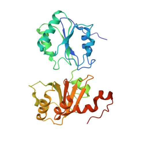
    1S4D

  • PubMed Abstract: 

    The crystallographic structure of the Pseudomonas denitrificans S-adenosyl-L-methionine-dependent uroporphyrinogen III methyltransferase (SUMT), which is encoded by the cobA gene, has been solved by molecular replacement to 2.7A resolution. SUMT is a branchpoint enzyme that plays a key role in the biosynthesis of modified tetrapyrroles by controlling flux to compounds such as vitamin B(12) and sirohaem, and catalysing the transformation of uroporphyrinogen III into precorrin-2. The overall topology of the enzyme is similar to that of the SUMT module of sirohaem synthase (CysG) and the cobalt-precorrin-4 methyltransferase CbiF and, as with the latter structures, SUMT has the product S-adenosyl-L-homocysteine bound in the crystal. The roles of a number of residues within the SUMT structure are discussed with respect to their conservation either across the broader family of cobalamin biosynthetic methyltransferases or within the sub-group of SUMT members. The D47N, L49A, F106A, T130A, Y183A and M184A variants of SUMT were generated by mutagenesis of the cobA gene, and tested for SAM binding and enzymatic activity. Of these variants, only D47N and L49A bound the co-substrate S-adenosyl-L-methionine. Consequently, all the mutants were severely restricted in their capacity to synthesise precorrin-2, although both the D47N and L49A variants produced significant quantities of precorrin-1, the monomethylated derivative of uroporphyrinogen III. The activity of these variants is interpreted with respect to the structure of the enzyme.


  • Organizational Affiliation
    • Structural Biology Laboratory, Department of Chemistry, University of York, Heslington, York YO10 5YW, UK.

Macromolecule Content 

  • Total Structure Weight: 358.45 kDa 
  • Atom Count: 23,427 
  • Modeled Residue Count: 3,075 
  • Deposited Residue Count: 3,360 
  • Unique protein chains: 1

Macromolecules

Find similar proteins by:|  3D Structure
Entity ID: 1
MoleculeChains  Sequence LengthOrganismDetailsImage
Uroporphyrin-III C-methyltransferase280Pseudomonas denitrificans (nom. rej.)Mutation(s): 0 
Gene Names: COBA
EC: 2.1.1.107
UniProt
Find proteins for P21631 (Sinorhizobium sp)
Explore P21631 
Go to UniProtKB:  P21631
Entity Groups
Sequence Clusters30% Identity50% Identity70% Identity90% Identity95% Identity100% Identity
UniProt GroupP21631
Sequence Annotations
Expand
Reference Sequence

Small Molecules

Ligands 2 Unique
IDChains Name / Formula / InChI Key2D Diagram3D Interactions
SAH

Query on SAH


Download:Ideal Coordinates CCD File
CA [auth G]
GA [auth H]
JA [auth I]
M [auth A]
MA [auth J]
CA [auth G],
GA [auth H],
JA [auth I],
M [auth A],
MA [auth J],
Q [auth B],
QA [auth K],
S [auth D],
TA [auth L],
V [auth E],
WA [auth M],
Z [auth F]
S-ADENOSYL-L-HOMOCYSTEINE
C14 H20 N6 O5 S
ZJUKTBDSGOFHSH-WFMPWKQPSA-N
GOL

Query on GOL


Download:Ideal Coordinates CCD File
AA [auth F]
BA [auth F]
DA [auth G]
EA [auth G]
FA [auth G]
AA [auth F],
BA [auth F],
DA [auth G],
EA [auth G],
FA [auth G],
HA [auth H],
IA [auth H],
KA [auth I],
LA [auth I],
N [auth A],
NA [auth J],
O [auth A],
OA [auth J],
P [auth A],
PA [auth J],
R [auth B],
RA [auth K],
SA [auth K],
T [auth D],
U [auth D],
UA [auth L],
VA [auth L],
W [auth E],
X [auth E],
XA [auth M],
Y [auth E]
GLYCEROL
C3 H8 O3
PEDCQBHIVMGVHV-UHFFFAOYSA-N

Experimental Data & Validation

Experimental Data

  • Method: X-RAY DIFFRACTION
  • Resolution: 2.70 Å
  • R-Value Free:  0.260 (Depositor), 0.249 (DCC) 
  • R-Value Work:  0.213 (Depositor) 
  • R-Value Observed: 0.215 (Depositor) 
Space Group: I 4
Unit Cell:
Length ( Å )Angle ( ˚ )
a = 218.097α = 90
b = 218.097β = 90
c = 190.341γ = 90
Software Package:
Software NamePurpose
REFMACrefinement
DENZOdata reduction
SCALEPACKdata scaling
AMoREphasing

Structure Validation

View Full Validation Report


Ligand Structure Quality Assessment 


Entry History 

Revision History  (Full details and data files)

  • Version 1.0: 2004-11-30
    Type: Initial release
  • Version 1.1: 2008-04-29
    Changes: Version format compliance
  • Version 1.2: 2011-07-13
    Changes: Advisory, Version format compliance
  • Version 1.3: 2023-10-25
    Changes: Data collection, Database references, Derived calculations, Refinement description