Download Mendeley
Structural insights into spectral tuning and retinal exchange in cone visual pigments.
Ohashi, S., Katayama, K., Kojima, A., Yang, X., Fukuda, M., Sacchetta, F., Suno, R., Sugita, Y., Nuemket, N., Kim, S., Kobayashi, K., Imai, H., Iwata, S., Nango, E., Kobayashi, T., Noda, T., Olivucci, M., Kato, H.E., Kandori, H.(2026) Science 392: eadz3996-eadz3996
- PubMed: 42348681 Search on PubMed
- DOI: https://doi.org/10.1126/science.adz3996
- Primary Citation Related Structures:
9UPM, 9UPN, 9UPO - PubMed Abstract:
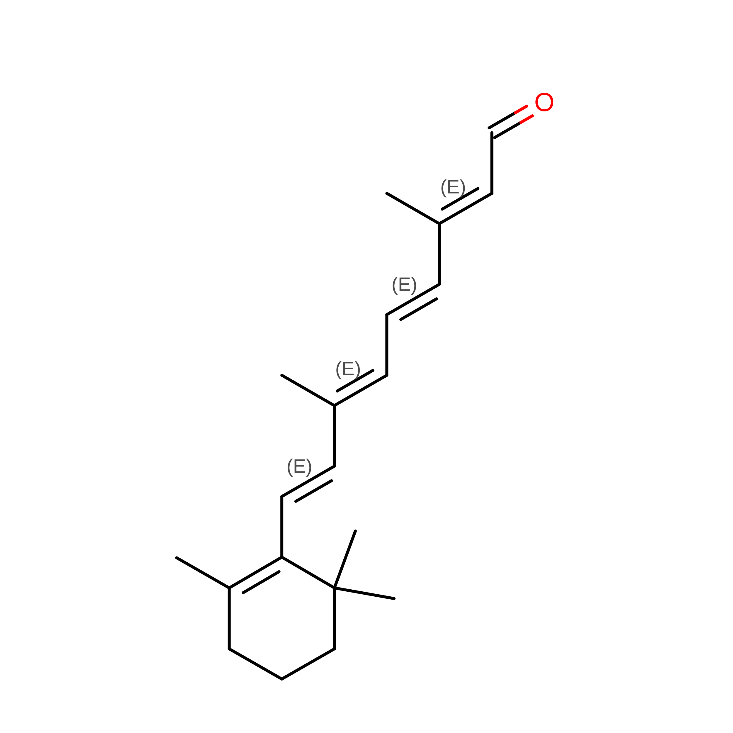
Color vision in catarrhine primates relies on red-, green-, and blue-sensitive cone pigments that share an 11- cis -retinal chromophore but differ in absorption maxima. Red and green pigments arose by recent gene duplication and differ at only a few residues. Here, we report cryo-electron microscopy structures of red and green cone pigments from the cynomolgus macaque ( Macaca fascicularis ) integrated with low-temperature vibrational spectroscopy and quantum mechanical and molecular mechanical modeling. The red-green spectral shift is dominated by threonine 285, the hydroxyl dipole of which modulates chromophore electrostatics, whereas steric effects appear modest. We also identified membrane-facing lateral openings in cone pigments but not in inactive rhodopsin. Comparisons with active-state structures suggest activation-dependent gating, and mutational and spectroscopic analyses support a role for this opening in retinal uptake and rapid pigment regeneration.
- Life Science and Applied Chemistry, Nagoya Institute of Technology, Nagoya, Japan.
Organizational Affiliation: