9UPM | pdb_00009upm

Cryo-EM structure of macaque red cone pigment with Q114N mutation


Experimental Data Snapshot

  • Method: ELECTRON MICROSCOPY
  • Resolution: 3.21 Å
  • Aggregation State: PARTICLE 
  • Reconstruction Method: SINGLE PARTICLE 

wwPDB Validation 3D Report Full Report

Validation slider image for 9UPM

This is version 1.1 of the entry. See complete history

Literature

Structural insights into spectral tuning and retinal exchange in cone visual pigments.

Ohashi, S.Katayama, K.Kojima, A.Yang, X.Fukuda, M.Sacchetta, F.Suno, R.Sugita, Y.Nuemket, N.Kim, S.Kobayashi, K.Imai, H.Iwata, S.Nango, E.Kobayashi, T.Noda, T.Olivucci, M.Kato, H.E.Kandori, H.

(2026) Science 392: eadz3996-eadz3996

  • DOI: https://doi.org/10.1126/science.adz3996
  • Primary Citation Related Structures: 
    9UPM, 9UPN, 9UPO

  • PubMed Abstract: 

    Color vision in catarrhine primates relies on red-, green-, and blue-sensitive cone pigments that share an 11- cis -retinal chromophore but differ in absorption maxima. Red and green pigments arose by recent gene duplication and differ at only a few residues. Here, we report cryo-electron microscopy structures of red and green cone pigments from the cynomolgus macaque ( Macaca fascicularis ) integrated with low-temperature vibrational spectroscopy and quantum mechanical and molecular mechanical modeling. The red-green spectral shift is dominated by threonine 285, the hydroxyl dipole of which modulates chromophore electrostatics, whereas steric effects appear modest. We also identified membrane-facing lateral openings in cone pigments but not in inactive rhodopsin. Comparisons with active-state structures suggest activation-dependent gating, and mutational and spectroscopic analyses support a role for this opening in retinal uptake and rapid pigment regeneration.


  • Organizational Affiliation
    • Life Science and Applied Chemistry, Nagoya Institute of Technology, Nagoya, Japan.

Macromolecule Content 

  • Total Structure Weight: 50.21 kDa 
  • Atom Count: 2,338 
  • Modeled Residue Count: 294 
  • Deposited Residue Count: 452 
  • Unique protein chains: 1

Macromolecules

Find similar proteins by:|  3D Structure
Entity ID: 1
MoleculeChains  Sequence LengthOrganismDetailsImage
LW opsin,Soluble cytochrome b562452Macaca fascicularisEscherichia coliMutation(s): 4 
UniProt
Find proteins for P0ABE7 (Escherichia coli)
Explore P0ABE7 
Go to UniProtKB:  P0ABE7
Find proteins for Q9XSX4 (Macaca fascicularis)
Explore Q9XSX4 
Go to UniProtKB:  Q9XSX4
Entity Groups
Sequence Clusters30% Identity50% Identity70% Identity90% Identity95% Identity100% Identity
UniProt GroupsP0ABE7Q9XSX4
Sequence Annotations
Expand
Reference Sequence

Small Molecules

Ligands 1 Unique
IDChains Name / Formula / InChI Key2D Diagram3D Interactions
RET
(Subject of Investigation/LOI)

Query on RET


Download:Ideal Coordinates CCD File
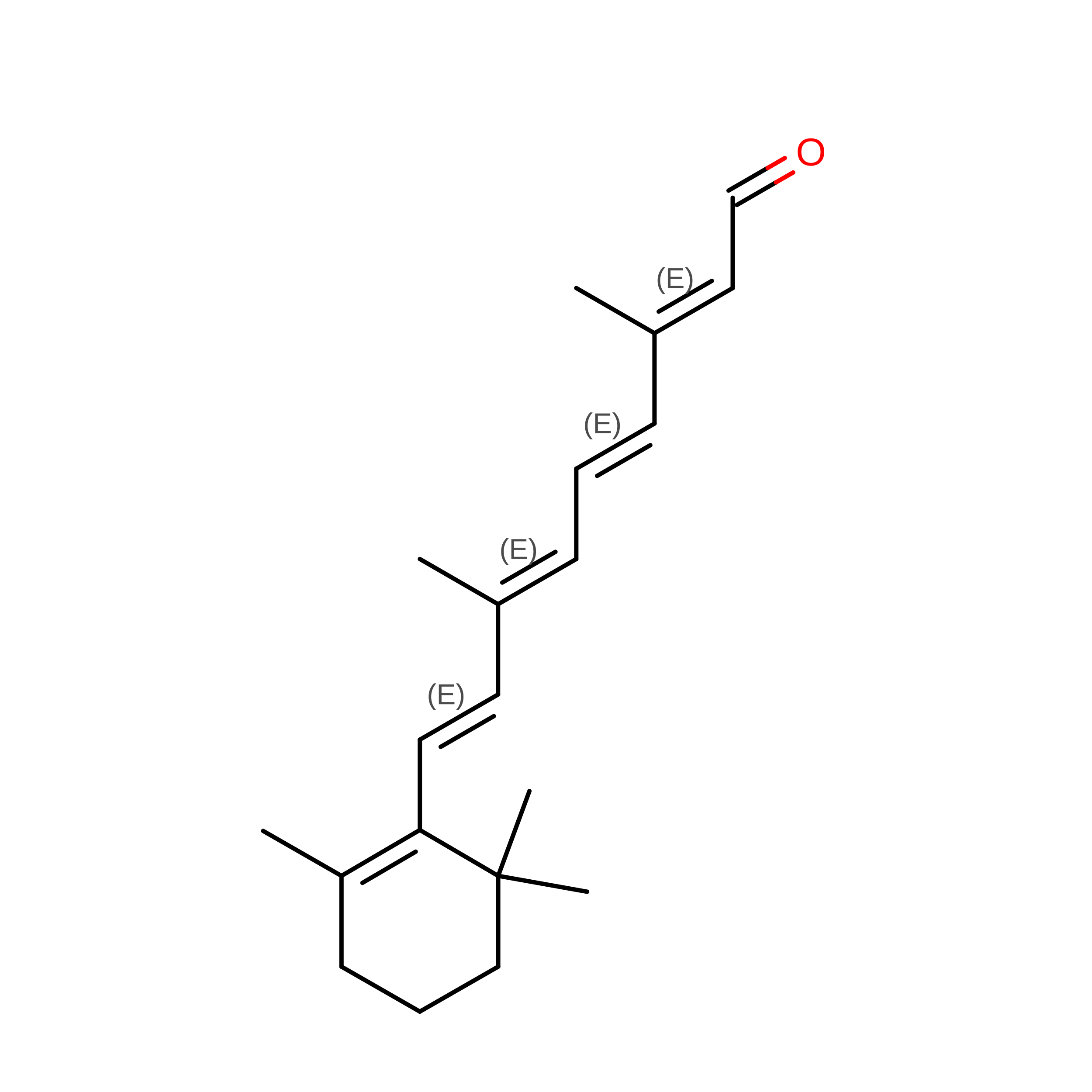
B [auth A]RETINAL
C20 H28 O
NCYCYZXNIZJOKI-OVSJKPMPSA-N

Experimental Data & Validation

Experimental Data

  • Method: ELECTRON MICROSCOPY
  • Resolution: 3.21 Å
  • Aggregation State: PARTICLE 
  • Reconstruction Method: SINGLE PARTICLE 

Structure Validation

View Full Validation Report


Entry History 

& Funding Information

Deposition Data


Funding OrganizationLocationGrant Number
Japan Science and TechnologyJapanJPMJFR204S
Japan Science and TechnologyJapanJPMJPR19G4
Japan Science and TechnologyJapanJPMJCR21P3
Japan Science and TechnologyJapanJPMJCR1753
Japan Science and TechnologyJapanJPMJCR23B1
Japan Science and TechnologyJapanJPMJAX222F
Japan Science and TechnologyJapanJPMJSP2112
Japan Science and TechnologyJapanJPMJPR24OF
Japan Society for the Promotion of Science (JSPS)Japan22H00400
Japan Society for the Promotion of Science (JSPS)Japan21H04969
Japan Society for the Promotion of Science (JSPS)Japan22K19265
Japan Society for the Promotion of Science (JSPS)JapanJP22H04742
Japan Society for the Promotion of Science (JSPS)JapanJP23K14142
Japan Society for the Promotion of Science (JSPS)JapanJP24H02262
Japan Society for the Promotion of Science (JSPS)Japan25K09525

Revision History  (Full details and data files)

  • Version 1.0: 2026-06-24
    Type: Initial release
  • Version 1.1: 2026-07-08
    Changes: Data collection, Database references