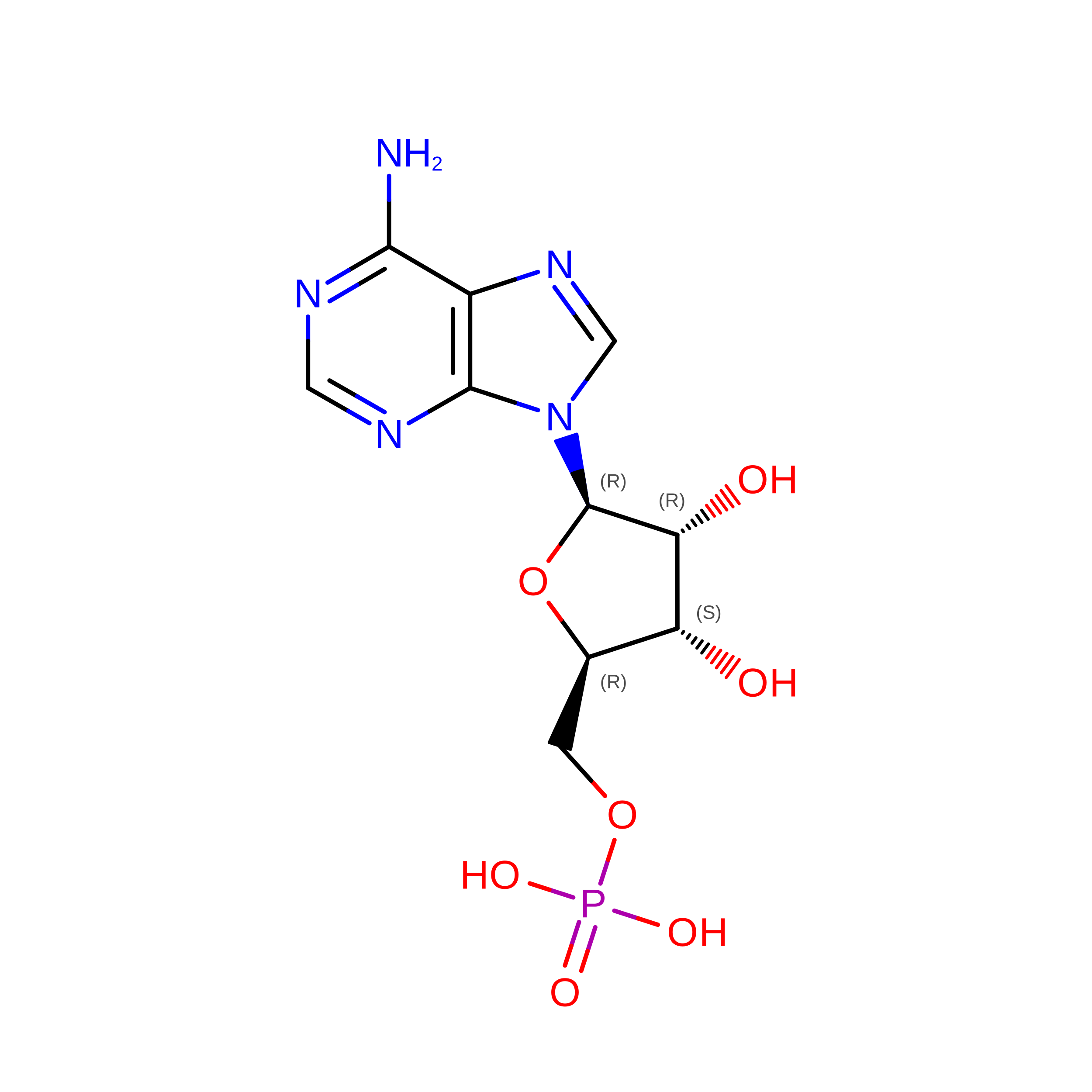
9YLF | pdb_00009ylf

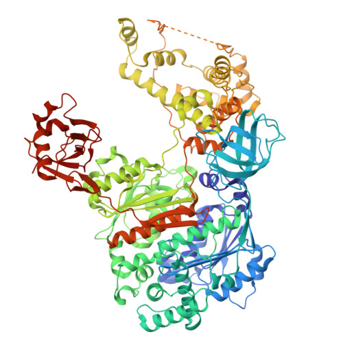
Cryo-EM structure of single-loaded human UBA6-UBE2Z/Ub(a) adenylate complex.


Experimental Data Snapshot

  • Method: ELECTRON MICROSCOPY
  • Resolution: 3.14 Å
  • Aggregation State: PARTICLE 
  • Reconstruction Method: SINGLE PARTICLE 

wwPDB Validation 3D Report Full Report

Validation slider image for 9YLF

This is version 1.0 of the entry. See complete history

Literature

Cryo-EM structures of UBA6 reveal mechanisms of E1-E2 specificity and dual FAT10/ubiquitin thioester transfer.

Nayak, D.Jia, L.Dos Santos Bury, P.Ruben, E.A.Shukla, A.Nayak, A.Stratton, C.M.Ebadi, P.Cho, H.Tumanova, A.A.Varughese, J.T.Yuan, L.Gao, F.Cano, K.E.Davies, C.Sung, P.Gack, M.U.Wasmuth, E.V.Olsen, S.K.

(2026) Nat Commun 17

  • DOI: https://doi.org/10.1038/s41467-026-69882-3
  • Primary Citation Related Structures: 
    9YKV, 9YKW, 9YLB, 9YLF

  • PubMed Abstract: 

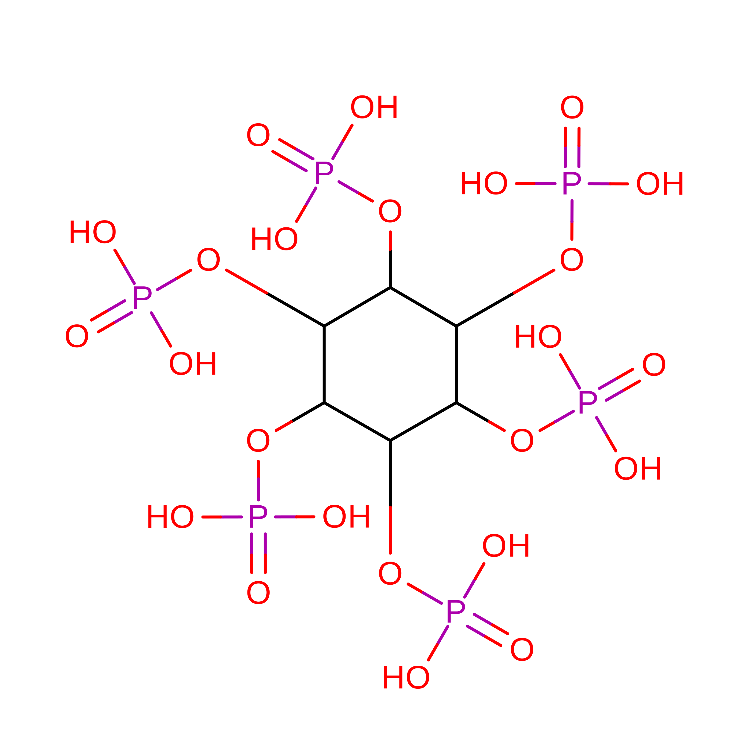
    UBA1 and UBA6 define parallel ubiquitin (Ub) activation systems that perform non-overlapping roles in Ub and ubiquitin-like protein (Ubl) signaling. Whereas UBA1 supports the canonical Ub pathway, UBA6 also activates the Ubl FAT10, linking Ub signaling to immune-regulated proteostasis. In addition to selective Ub/Ubl activation, UBA1 and UBA6 engage distinct sets of E2s, yet how these enzymes achieve selective E2 engagement has remained unclear. Using chemical trapping and high-resolution cryo-EM, we determine four structures of UBA6-E2 complexes representing the thioester-transfer step with either FAT10 or Ub, revealing how this E1 distinguishes its cognate partners. UBA6 achieves E2 specificity through coordinated contributions of the UFD and SCCH domains, a dual-domain mechanism that contrasts with the UFD-dominated selectivity of UBA1. The structures further show that an existing inositol hexakisphosphate (InsP₆)-binding site, unique to UBA6, stabilizes an expanded SCCH cleft that pre-organizes the enzyme for selective engagement of UBA6-specific E2s. These findings define principles for E1-E2 recognition and identify InsP₆ as a cofactor shaping specificity within the Ub-like conjugation network.


  • Organizational Affiliation
    • Department of Biochemistry & Structural Biology and Greehey Children's Cancer Research Institute, The University of Texas Health Science Center at San Antonio, San Antonio, TX, USA.

Macromolecule Content 

  • Total Structure Weight: 153.88 kDa 
  • Atom Count: 10,302 
  • Modeled Residue Count: 1,291 
  • Deposited Residue Count: 1,354 
  • Unique protein chains: 3

Macromolecules

Find similar proteins by:|  3D Structure
Entity ID: 1
MoleculeChains  Sequence LengthOrganismDetailsImage
Ubiquitin-like modifier-activating enzyme 61,016Homo sapiensMutation(s): 0 
Gene Names: UBA6MOP4UBE1L2
EC: 6.2.1.45
UniProt & NIH Common Fund Data Resources
Find proteins for A0AVT1 (Homo sapiens)
Explore A0AVT1 
Go to UniProtKB:  A0AVT1
PHAROS:  A0AVT1
GTEx:  ENSG00000033178 
Entity Groups
Sequence Clusters30% Identity50% Identity70% Identity90% Identity95% Identity100% Identity
UniProt GroupA0AVT1
Sequence Annotations
Expand
Reference Sequence
Find similar proteins by:|  3D Structure
Entity ID: 2
MoleculeChains  Sequence LengthOrganismDetailsImage
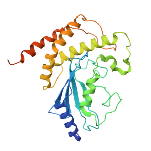
Ubiquitin-conjugating enzyme E2 ZB [auth C]262Homo sapiensMutation(s): 4 
Gene Names: UBE2ZHOYS7
EC: 2.3.2.23
UniProt & NIH Common Fund Data Resources
Find proteins for Q9H832 (Homo sapiens)
Explore Q9H832 
Go to UniProtKB:  Q9H832
PHAROS:  Q9H832
GTEx:  ENSG00000159202 
Entity Groups
Sequence Clusters30% Identity50% Identity70% Identity90% Identity95% Identity100% Identity
UniProt GroupQ9H832
Sequence Annotations
Expand
Reference Sequence
Find similar proteins by:|  3D Structure
Entity ID: 3
MoleculeChains  Sequence LengthOrganismDetailsImage
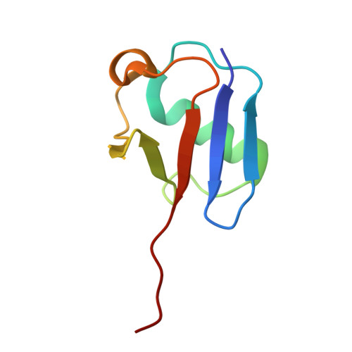
UbiquitinC [auth D]76Homo sapiensMutation(s): 0 
Gene Names: UBC
UniProt & NIH Common Fund Data Resources
Find proteins for P0CG48 (Homo sapiens)
Explore P0CG48 
Go to UniProtKB:  P0CG48
PHAROS:  P0CG48
GTEx:  ENSG00000150991 
Entity Groups
Sequence Clusters30% Identity50% Identity70% Identity90% Identity95% Identity100% Identity
UniProt GroupP0CG48
Sequence Annotations
Expand
Reference Sequence

Experimental Data & Validation

Experimental Data

  • Method: ELECTRON MICROSCOPY
  • Resolution: 3.14 Å
  • Aggregation State: PARTICLE 
  • Reconstruction Method: SINGLE PARTICLE 
EM Software:
TaskSoftware PackageVersion
MODEL REFINEMENTPHENIX1.20.1_4487
RECONSTRUCTIONcryoSPARC

Structure Validation

View Full Validation Report


Entry History 

& Funding Information

Deposition Data


Funding OrganizationLocationGrant Number
National Institutes of Health/National Institute of General Medical Sciences (NIH/NIGMS)United States--

Revision History  (Full details and data files)

  • Version 1.0: 2026-08-12
    Type: Initial release