9YKW | pdb_00009ykw

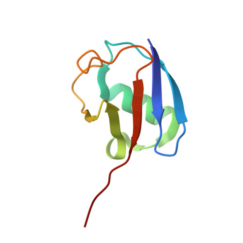
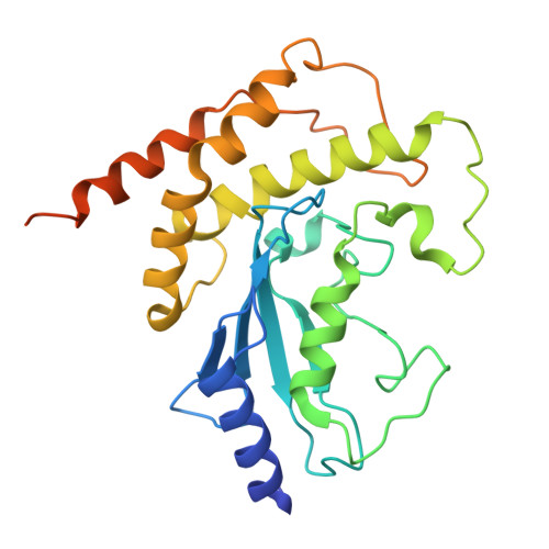
Cryo-EM structure of double-loaded human UBA6-UBE2Z-Ub(t)/Ub(a) thioester mimetic complex.


Experimental Data Snapshot

  • Method: ELECTRON MICROSCOPY
  • Resolution: 3.86 Å
  • Aggregation State: PARTICLE 
  • Reconstruction Method: SINGLE PARTICLE 

wwPDB Validation 3D Report Full Report

Validation slider image for 9YKW

This is version 1.0 of the entry. See complete history. 

Literature

Cryo-EM structures of UBA6 reveal mechanisms of E1-E2 specificity and dual FAT10/ubiquitin thioester transfer.

Nayak, D., Jia, L., Dos Santos Bury, P., Ruben, E.A., Shukla, A., Nayak, A., Stratton, C.M., Ebadi, P., Cho, H., Tumanova, A.A., Varughese, J.T., Yuan, L., Gao, F., Cano, K.E., Davies, C., Sung, P., Gack, M.U., Wasmuth, E.V., Olsen, S.K.

(2026) Nat Commun 17

  • DOI: https://doi.org/10.1038/s41467-026-69882-3
  • Primary Citation Related Structures: 
    9YKV, 9YKW, 9YLB, 9YLF

  • PubMed Abstract: 

    UBA1 and UBA6 define parallel ubiquitin (Ub) activation systems that perform non-overlapping roles in Ub and ubiquitin-like protein (Ubl) signaling. Whereas UBA1 supports the canonical Ub pathway, UBA6 also activates the Ubl FAT10, linking Ub signaling to immune-regulated proteostasis. In addition to selective Ub/Ubl activation, UBA1 and UBA6 engage distinct sets of E2s, yet how these enzymes achieve selective E2 engagement has remained unclear. Using chemical trapping and high-resolution cryo-EM, we determine four structures of UBA6-E2 complexes representing the thioester-transfer step with either FAT10 or Ub, revealing how this E1 distinguishes its cognate partners. UBA6 achieves E2 specificity through coordinated contributions of the UFD and SCCH domains, a dual-domain mechanism that contrasts with the UFD-dominated selectivity of UBA1. The structures further show that an existing inositol hexakisphosphate (InsP₆)-binding site, unique to UBA6, stabilizes an expanded SCCH cleft that pre-organizes the enzyme for selective engagement of UBA6-specific E2s. These findings define principles for E1-E2 recognition and identify InsP₆ as a cofactor shaping specificity within the Ub-like conjugation network.


  • Organizational Affiliation: 
    • Department of Biochemistry & Structural Biology and Greehey Children's Cancer Research Institute, The University of Texas Health Science Center at San Antonio, San Antonio, TX, USA.

Macromolecule Content 

  • Total Structure Weight: 162.2 kDa 
  • Atom Count: 10,794 
  • Modeled Residue Count: 1,354 
  • Deposited Residue Count: 1,430 
  • Unique protein chains: 3

Macromolecules

Find similar proteins by:|  3D Structure
Entity ID: 1
MoleculeChains  Sequence LengthOrganismDetailsImage
Ubiquitin-like modifier-activating enzyme 61,016Homo sapiensMutation(s): 0 
Gene Names: UBA6, MOP4, UBE1L2
EC: 6.2.1.45
UniProt & NIH Common Fund Data Resources
Find proteins for A0AVT1 (Homo sapiens)
Explore A0AVT1 
Go to UniProtKB:  A0AVT1
PHAROS:  A0AVT1
GTEx:  ENSG00000033178 
Entity Groups
Sequence Clusters30% Identity50% Identity70% Identity90% Identity95% Identity100% Identity
UniProt GroupA0AVT1
Sequence Annotations
Expand
Reference Sequence
Find similar proteins by:|  3D Structure
Entity ID: 2
MoleculeChains  Sequence LengthOrganismDetailsImage
Ubiquitin
B, D
76Homo sapiensMutation(s): 0 
Gene Names: UBC
UniProt & NIH Common Fund Data Resources
Find proteins for P0CG48 (Homo sapiens)
Explore P0CG48 
Go to UniProtKB:  P0CG48
PHAROS:  P0CG48
GTEx:  ENSG00000150991 
Entity Groups
Sequence Clusters30% Identity50% Identity70% Identity90% Identity95% Identity100% Identity
UniProt GroupP0CG48
Sequence Annotations
Expand
Reference Sequence
Find similar proteins by:|  3D Structure
Entity ID: 3
MoleculeChains  Sequence LengthOrganismDetailsImage
Ubiquitin-conjugating enzyme E2 Z262Homo sapiensMutation(s): 5 
Gene Names: UBE2Z, HOYS7
EC: 2.3.2.23
UniProt & NIH Common Fund Data Resources
Find proteins for Q9H832 (Homo sapiens)
Explore Q9H832 
Go to UniProtKB:  Q9H832
PHAROS:  Q9H832
GTEx:  ENSG00000159202 
Entity Groups
Sequence Clusters30% Identity50% Identity70% Identity90% Identity95% Identity100% Identity
UniProt GroupQ9H832
Sequence Annotations
Expand
Reference Sequence

Small Molecules

Ligands 1 Unique
IDChains Name / Formula / InChI Key2D Diagram3D Interactions
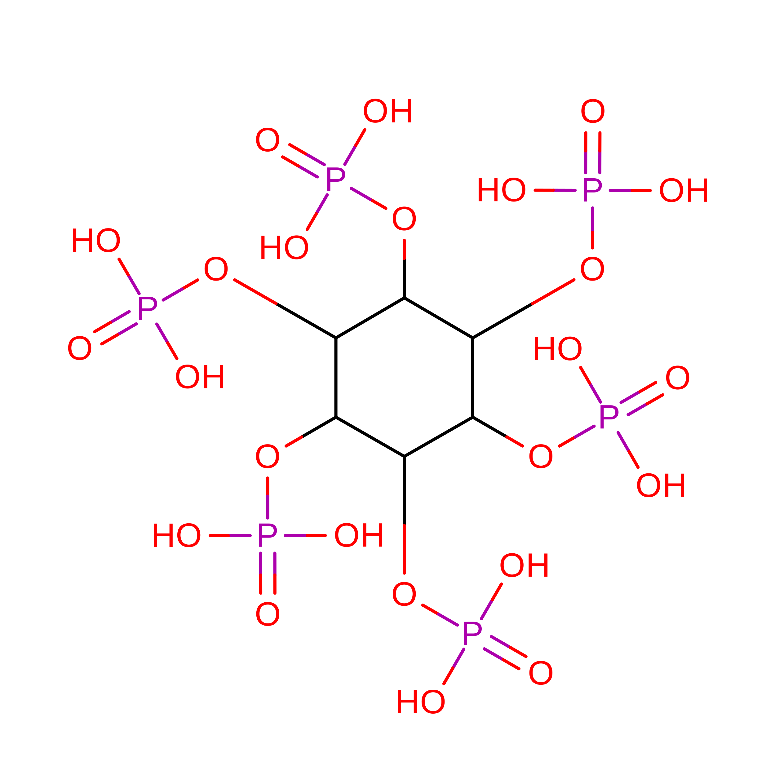
IHP
(Subject of Investigation/LOI)

Query on IHP


Download:Ideal Coordinates CCD File
E [auth A]INOSITOL HEXAKISPHOSPHATE
C6 H18 O24 P6
IMQLKJBTEOYOSI-GPIVLXJGSA-N

Experimental Data & Validation

Experimental Data

  • Method: ELECTRON MICROSCOPY
  • Resolution: 3.86 Å
  • Aggregation State: PARTICLE 
  • Reconstruction Method: SINGLE PARTICLE 
EM Software:
TaskSoftware PackageVersion
MODEL REFINEMENTPHENIX1.20.1_4487
RECONSTRUCTIONcryoSPARC

Structure Validation

View Full Validation Report


Entry History 

& Funding Information

Deposition Data


Funding OrganizationLocationGrant Number
National Institutes of Health/National Institute of General Medical Sciences (NIH/NIGMS)United States--

Revision History  (Full details and data files)

  • Version 1.0: 2026-08-12
    Type: Initial release