9UAL | pdb_00009ual

Crystal structure of Trimucrin


Experimental Data Snapshot

  • Method: X-RAY DIFFRACTION
  • Resolution: 1.68 Å
  • R-Value Free: 
    0.217 (Depositor), 0.225 (DCC) 
  • R-Value Work: 
    0.174 (Depositor), 0.184 (DCC) 

Starting Model: experimental
View more details

wwPDB Validation 3D Report Full Report

Validation slider image for 9UAL

This is version 1.2 of the entry. See complete history

Literature

Structural basis for the differential recognition of integrin alpha v beta 3 by rhodostomin and trimucrin.

Wang, Y.T.Chang, Y.T.Huang, C.H.Liau, C.T.Chen, C.Y.Chuang, W.J.

(2026) Commun Biol 9

  • DOI: https://doi.org/10.1038/s42003-026-10139-6
  • Primary Citation Related Structures: 
    9IUJ, 9LT3, 9UAL

  • PubMed Abstract: 

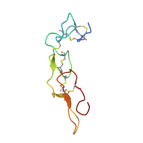
    Rhodostomin (Rho) and Trimucrin (Tmu) are RGD-containing disintegrins that inhibit integrins more effectively than short RGD peptides. They differ in their linker, RGD loop, and C-terminal sequences. We determined the X-ray structure of Tmu and the cryo-EM structures of integrin αvβ3 in complex with both disintegrins. Structural analysis revealed subtle differences in binding, with both adopting a rigid backbone conformation and interacting with integrin through three cooperative binding sites. Besides the conserved RGD interface, Tmu features a cluster of basic residues in its linker, while Rho has distinct C-terminal interactions. Disintegrin binding stabilizes αvβ3 in an extended-open conformation, while the β3-Y110 residue is essential for maintaining the bent state without ligands. These findings enhance our understanding of integrin recognition and inform the development of integrin-targeted therapeutics for anti-angiogenic, anti-tumor, and anti-inflammatory applications.


  • Organizational Affiliation
    • Institute of Basic Medical Sciences, National Cheng Kung University College of Medicine, Tainan, Taiwan, ROC.

Macromolecule Content 

  • Total Structure Weight: 16.46 kDa 
  • Atom Count: 1,170 
  • Modeled Residue Count: 140 
  • Deposited Residue Count: 146 
  • Unique protein chains: 1

Macromolecules

Find similar proteins by:|  3D Structure
Entity ID: 1
MoleculeChains  Sequence LengthOrganismDetailsImage
Zinc metalloproteinase/disintegrin PMMP-2
A, B
73Protobothrops mucrosquamatusMutation(s): 0 
UniProt
Find proteins for E9NW27 (Protobothrops mucrosquamatus)
Explore E9NW27 
Go to UniProtKB:  E9NW27
Entity Groups
Sequence Clusters30% Identity50% Identity70% Identity90% Identity95% Identity100% Identity
UniProt GroupE9NW27
Sequence Annotations
Expand
Reference Sequence

Small Molecules

Ligands 2 Unique
IDChains Name / Formula / InChI Key2D Diagram3D Interactions
ZN

Query on ZN


Download:Ideal Coordinates CCD File
C [auth A]
D [auth A]
H [auth B]
I [auth B]
J [auth B]
C [auth A],
D [auth A],
H [auth B],
I [auth B],
J [auth B],
K [auth B]
ZINC ION
Zn
PTFCDOFLOPIGGS-UHFFFAOYSA-N
ACT

Query on ACT


Download:Ideal Coordinates CCD File
E [auth A],
F [auth A],
G [auth A],
L [auth B],
M [auth B]
ACETATE ION
C2 H3 O2
QTBSBXVTEAMEQO-UHFFFAOYSA-M

Experimental Data & Validation

Experimental Data

  • Method: X-RAY DIFFRACTION
  • Resolution: 1.68 Å
  • R-Value Free:  0.217 (Depositor), 0.225 (DCC) 
  • R-Value Work:  0.174 (Depositor), 0.184 (DCC) 
Space Group: P 1 21 1
Unit Cell:
Length ( Å )Angle ( ˚ )
a = 26.954α = 90
b = 58.397β = 90.266
c = 42.871γ = 90
Software Package:
Software NamePurpose
REFMACrefinement
XDSdata reduction
XDSdata scaling
PHASERphasing

Structure Validation

View Full Validation Report


Entry History 

& Funding Information

Deposition Data


Funding OrganizationLocationGrant Number
Ministry of Science and Technology (MoST, Taiwan)Taiwan--

Revision History  (Full details and data files)

  • Version 1.0: 2026-04-08
    Type: Initial release
  • Version 1.1: 2026-04-15
    Changes: Database references
  • Version 1.2: 2026-05-20
    Changes: Database references