9SL1 | pdb_00009sl1

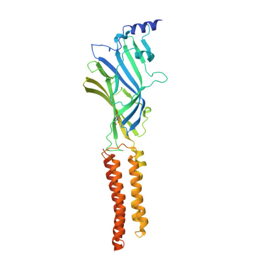
GABA-A a4b3d receptor in complex with GABA, Nb24 and Mb30


Experimental Data Snapshot

  • Method: ELECTRON MICROSCOPY
  • Resolution: 2.68 Å
  • Aggregation State: PARTICLE 
  • Reconstruction Method: SINGLE PARTICLE 

Starting Model: experimental
View more details

wwPDB Validation 3D Report Full Report

Validation slider image for 9SL1

This is version 1.1 of the entry. See complete history. 

Literature

Molecular arrangement and modulation of extrasynaptic alpha beta delta GABA A receptors.

Nestorow, S.A., Chen, W.N., Bertrand, D., Wahid, A.A., Wyatt, C.E., Mortensen, M., Clairfeuille, T., Reutlinger, M., Friz, G., Pardon, E., Steyaert, J., Ladds, G., Cecere, G., Smart, T.G., Hernandez, M.C., Joedicke, L., Miller, P.S.

(2026) Nat Commun 17

  • DOI: https://doi.org/10.1038/s41467-026-76879-5
  • Primary Citation Related Structures: 
    9SL1, 9SLD, 9SLM

  • PubMed Abstract: 

    γ-aminobutyric acid Type-A (GABA A ) αβδ receptors regulate neuronal excitability and contribute to sleep, mood and motor coordination. However, the molecular arrangements of these receptors and the basis of GABA activation and δ-selective drug modulation remain unclear. We solve cryo-EM structures of α4β3δ receptors in an α-β-α-β-δ arrangement that contains two GABA binding pockets. GABA binding supports a classical β-subunit tilt and an outward configuration of the β-subunit M2-M3 loops. However, the δ subunit M2-M3 loop is orientated inward and the 9' activation gate in the pore is closed, consistent with the low efficacy for these receptors. Addition of a δ-selective positive allosteric modulator (PAM), DS2-Me, reveals binding to an α-δ pocket that causes the 9' activation gate to open. In this work we provide key insights into the stoichiometry, arrangement and molecular modes of GABA activation and δ-selective modulation of these critical regulators of neuronal excitability.


  • Organizational Affiliation: 
    • Department of Pharmacology, University of Cambridge, Cambridge, UK.

Macromolecule Content 

  • Total Structure Weight: 299.33 kDa 
  • Atom Count: 17,537 
  • Modeled Residue Count: 2,151 
  • Deposited Residue Count: 2,602 
  • Unique protein chains: 5

Macromolecules

Find similar proteins by:|  3D Structure
Entity ID: 1
MoleculeChains  Sequence LengthOrganismDetailsImage
Gamma-aminobutyric acid receptor subunit alpha-4
A, C
364Homo sapiensMutation(s): 1 
Gene Names: GABRA4
UniProt & NIH Common Fund Data Resources
Find proteins for P48169 (Homo sapiens)
Explore P48169 
Go to UniProtKB:  P48169
PHAROS:  P48169
GTEx:  ENSG00000109158 
Entity Groups
Sequence Clusters30% Identity50% Identity70% Identity90% Identity95% Identity100% Identity
UniProt GroupP48169
Glycosylation
Glycosylation Sites: 1Go to GlyGen: P48169-1
Sequence Annotations
Expand
Reference Sequence
Find similar proteins by:|  3D Structure
Entity ID: 2
MoleculeChains  Sequence LengthOrganismDetailsImage
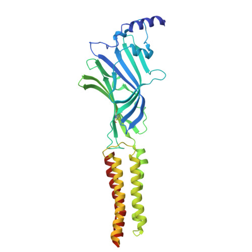
Gamma-aminobutyric acid receptor subunit beta-3,Soluble cytochrome b562
B, D
451Homo sapiens, Escherichia coliMutation(s): 3 
Gene Names: GABRB3, cybC
UniProt & NIH Common Fund Data Resources
Find proteins for P28472 (Homo sapiens)
Explore P28472 
Go to UniProtKB:  P28472
PHAROS:  P28472
GTEx:  ENSG00000166206 
Find proteins for P0ABE7 (Escherichia coli)
Explore P0ABE7 
Go to UniProtKB:  P0ABE7
Entity Groups
Sequence Clusters30% Identity50% Identity70% Identity90% Identity95% Identity100% Identity
UniProt GroupsP0ABE7P28472
Glycosylation
Glycosylation Sites: 2Go to GlyGen: P28472-1
Sequence Annotations
Expand
Reference Sequence
Find similar proteins by:|  3D Structure
Entity ID: 3
MoleculeChains  Sequence LengthOrganismDetailsImage
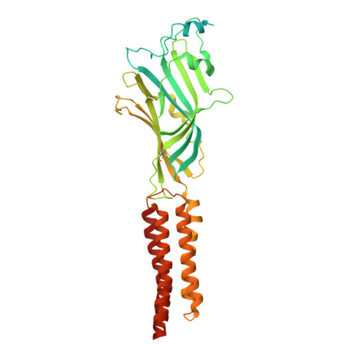
GABA-A receptor SBP-delta-glvi,Gamma-aminobutyric acid receptor subunit delta,Gamma-aminobutyric acid receptor subunit delta430Homo sapiensMutation(s): 0 
UniProt & NIH Common Fund Data Resources
Find proteins for O14764 (Homo sapiens)
Explore O14764 
Go to UniProtKB:  O14764
PHAROS:  O14764
GTEx:  ENSG00000187730 
Entity Groups
Sequence Clusters30% Identity50% Identity70% Identity90% Identity95% Identity100% Identity
UniProt GroupO14764
Glycosylation
Glycosylation Sites: 1Go to GlyGen: O14764-1
Sequence Annotations
Expand
Reference Sequence
Find similar proteins by:|  3D Structure
Entity ID: 4
MoleculeChains  Sequence LengthOrganismDetailsImage
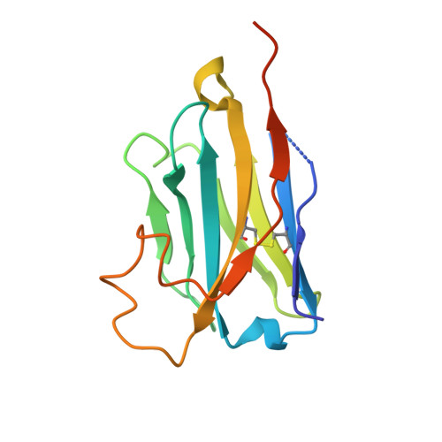
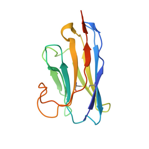
Megabody Mb30
F, H
138Lama glamaMutation(s): 0 
Entity Groups
Sequence Clusters30% Identity50% Identity70% Identity90% Identity95% Identity100% Identity
Sequence Annotations
Expand
Reference Sequence
Find similar proteins by:|  3D Structure
Entity ID: 5
MoleculeChains  Sequence LengthOrganismDetailsImage
Nanobody Nb24
G, I
133Lama glamaMutation(s): 0 
Entity Groups
Sequence Clusters30% Identity50% Identity70% Identity90% Identity95% Identity100% Identity
Sequence Annotations
Expand
Reference Sequence

Oligosaccharides

Help  
Entity ID: 6
MoleculeChains Length2D Diagram GlycosylationD Interactions
beta-D-mannopyranose-(1-4)-2-acetamido-2-deoxy-beta-D-glucopyranose-(1-3)-2-acetamido-2-deoxy-beta-D-glucopyranoseJ [auth Q]3N-Glycosylation
Glycosylation Resources
GlyTouCan: G53081KU
GlyCosmos: G53081KU
GlyGen: G53081KU
Entity ID: 7
MoleculeChains Length2D Diagram GlycosylationD Interactions
beta-D-mannopyranose-(1-3)-2-acetamido-2-deoxy-beta-D-glucopyranose-(1-3)-2-acetamido-2-deoxy-beta-D-glucopyranoseK [auth R]3N-Glycosylation

Small Molecules

Ligands 4 Unique
IDChains Name / Formula / InChI Key2D Diagram3D Interactions
PX6

Query on PX6


Download:Ideal Coordinates CCD File
N [auth A]1,2-DIPALMITOYL-SN-GLYCERO-3-PHOSPHATE
C35 H68 O8 P
PORPENFLTBBHSG-MGBGTMOVSA-M
NAG

Query on NAG


Download:Ideal Coordinates CCD File
L [auth A]
M [auth A]
Q [auth B]
R [auth B]
T [auth C]
L [auth A],
M [auth A],
Q [auth B],
R [auth B],
T [auth C],
V [auth D],
X [auth E]
2-acetamido-2-deoxy-beta-D-glucopyranose
C8 H15 N O6
OVRNDRQMDRJTHS-FMDGEEDCSA-N
D10

Query on D10


Download:Ideal Coordinates CCD File
O [auth A],
S [auth B],
W [auth D],
Y [auth E]
DECANE
C10 H22
DIOQZVSQGTUSAI-UHFFFAOYSA-N
ABU
(Subject of Investigation/LOI)

Query on ABU


Download:Ideal Coordinates CCD File
P [auth B],
U [auth D]
GAMMA-AMINO-BUTANOIC ACID
C4 H9 N O2
BTCSSZJGUNDROE-UHFFFAOYSA-N

Experimental Data & Validation

Experimental Data

  • Method: ELECTRON MICROSCOPY
  • Resolution: 2.68 Å
  • Aggregation State: PARTICLE 
  • Reconstruction Method: SINGLE PARTICLE 
EM Software:
TaskSoftware PackageVersion
MODEL REFINEMENTPHENIX1.20.1_4487
RECONSTRUCTIONcryoSPARC4.4.1 to 4.7.1

Structure Validation

View Full Validation Report


Entry History 

& Funding Information

Deposition Data


Funding OrganizationLocationGrant Number
Biotechnology and Biological Sciences Research Council (BBSRC)United KingdomBB/M024709/1
F. Hoffmann-La Roche LTDSwitzerland--

Revision History  (Full details and data files)

  • Version 1.0: 2026-09-02
    Type: Initial release
  • Version 1.1: 2026-09-30
    Changes: Data collection, Database references