9RBU | pdb_00009rbu

Cryo-ET structure of full-length membrane-bound EHD2 complex


Experimental Data Snapshot

  • Method: ELECTRON MICROSCOPY
  • Resolution: 6.70 Å
  • Aggregation State: FILAMENT 
  • Reconstruction Method: SUBTOMOGRAM AVERAGING 

Starting Model: experimental
View more details

wwPDB Validation 3D Report Full Report

Validation slider image for 9RBU

This is version 1.1 of the entry. See complete history

Literature

Structures of EHD2 filaments on curved membranes provide a model for caveolar neck stabilization.

Vazquez-Sarandeses, E.Mikirtumov, V.Noel, J.K.Kudryashev, M.Daumke, O.

(2026) Nat Commun 17

  • DOI: https://doi.org/10.1038/s41467-026-76288-8
  • Primary Citation Related Structures: 
    9RBU, 9RC1

  • PubMed Abstract: 

    Caveolae are flask-shaped invaginations of the plasma membrane serving critical functions in mechano-protection and signal transduction. Caveolar dynamics, such as caveolar movement within the plasma membrane or endocytosis, relies on precise shaping of the highly curved caveolar necks. The dynamin-like EHD2 ATPase is proposed to oligomerize around the caveolar neck, but its detailed molecular action is poorly understood. Here, we employ cryo-electron tomography to elucidate structures of ring-like EHD2 filaments on tubulated liposomes. EHD2 forms highly curved membrane scaffolds which stabilize a tubular membrane geometry with undulations along the tube's axis, resembling caveolar neck architecture. An amino-terminal sequence facilitates this geometry by acting as a spacer between adjacent filaments. Moreover, in endothelial cells lacking EHD2, caveolar necks become narrower and more elongated. Our structural work provides the molecular framework for understanding EHD2 scaffold formation and its cellular function in caveolar dynamics.


  • Organizational Affiliation
    • Structural Biology, Max Delbrück Center for Molecular Medicine, Berlin, Germany.

Macromolecule Content 

  • Total Structure Weight: 247.24 kDa 
  • Atom Count: 16,024 
  • Modeled Residue Count: 2,028 
  • Deposited Residue Count: 2,172 
  • Unique protein chains: 1

Macromolecules

Find similar proteins by:|  3D Structure
Entity ID: 1
MoleculeChains  Sequence LengthOrganismDetailsImage
EH domain-containing protein 2
A, B, C, D
543Mus musculusMutation(s): 0 
Gene Names: Ehd2
UniProt
Find proteins for Q8BH64 (Mus musculus)
Explore Q8BH64 
Go to UniProtKB:  Q8BH64
Entity Groups
Sequence Clusters30% Identity50% Identity70% Identity90% Identity95% Identity100% Identity
UniProt GroupQ8BH64
Sequence Annotations
Expand
Reference Sequence

Small Molecules

Ligands 2 Unique
IDChains Name / Formula / InChI Key2D Diagram3D Interactions
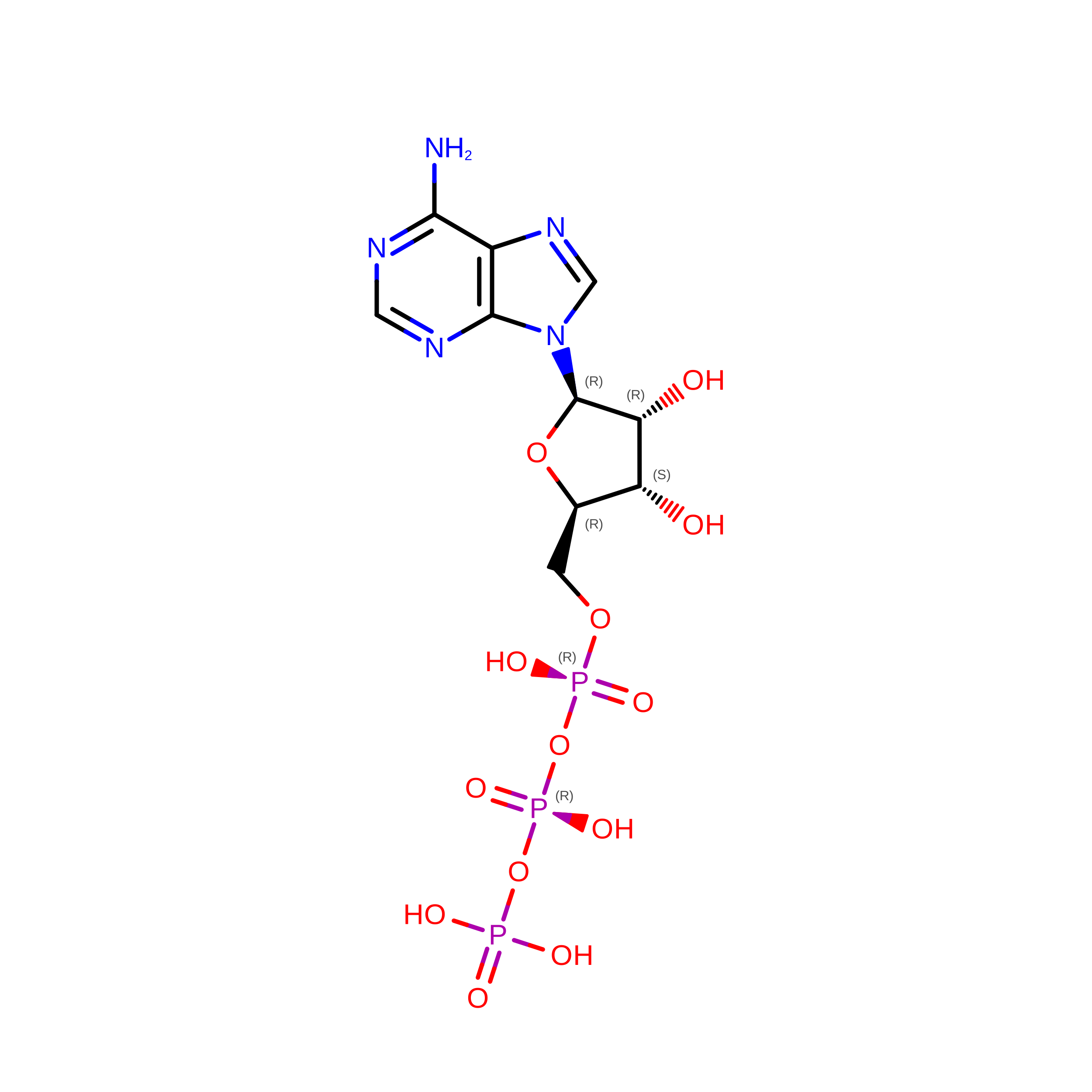
ATP
(Subject of Investigation/LOI)

Query on ATP


Download:Ideal Coordinates CCD File
E [auth A],
G [auth B],
I [auth C],
K [auth D]
ADENOSINE-5'-TRIPHOSPHATE
C10 H16 N5 O13 P3
ZKHQWZAMYRWXGA-KQYNXXCUSA-N
MG
(Subject of Investigation/LOI)

Query on MG


Download:Ideal Coordinates CCD File
F [auth A],
H [auth B],
J [auth C],
L [auth D]
MAGNESIUM ION
Mg
JLVVSXFLKOJNIY-UHFFFAOYSA-N

Experimental Data & Validation

Experimental Data

  • Method: ELECTRON MICROSCOPY
  • Resolution: 6.70 Å
  • Aggregation State: FILAMENT 
  • Reconstruction Method: SUBTOMOGRAM AVERAGING 
EM Software:
TaskSoftware PackageVersion
RECONSTRUCTIONRELION4
MODEL REFINEMENTPHENIX

Structure Validation

View Full Validation Report


Entry History 

& Funding Information

Deposition Data


Funding OrganizationLocationGrant Number
Helmholtz AssociationGermany--

Revision History  (Full details and data files)

  • Version 1.0: 2026-07-08
    Type: Initial release
  • Version 1.1: 2026-08-19
    Changes: Data collection, Database references