9O6N | pdb_00009o6n

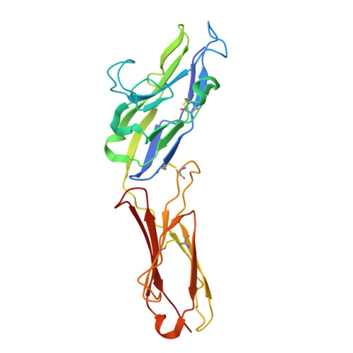
Structure of Siglec-10


Experimental Data Snapshot

  • Method: X-RAY DIFFRACTION
  • Resolution: 2.98 Å
  • R-Value Free: 
    0.259 (Depositor), 0.259 (DCC) 
  • R-Value Work: 
    0.212 (Depositor), 0.218 (DCC) 
  • R-Value Observed: 
    0.217 (Depositor) 

Starting Model: in silico
View more details

wwPDB Validation 3D Report Full Report

Validation slider image for 9O6N

Ligand Structure Quality Assessment 


This is version 1.1 of the entry. See complete history

Literature

Structural basis for sialoglycan recognition by the immune inhibitory receptor Siglec-10.

Medina, E.Mason, C.Tran, T.H.Julia, E.P.Ming, Q.Taibi, L.M.Hwu, P.Maute, R.L.Burg, J.S.Luca, V.C.

(2026) Structure 34: 768-777.e5

  • DOI: https://doi.org/10.1016/j.str.2026.01.018
  • Primary Citation Related Structures: 
    9O6J, 9O6N, 9O6O

  • PubMed Abstract: 

    Sialic acid-binding immunoglobulin-like lectin 10 (Siglec-10) inhibits immune cell function by sensing the presence of sialylated glycoproteins. Here, we determined structures of Siglec-10 bound to sialyllactose (SL) ligands to visualize the molecular recognition events underlying Siglec-10 signaling. The structures reveal that domain 1 (D1) of Siglec-10 engages SL using a non-conserved, selectivity determining CC' loop. Siglec-10 binds α2,3- and α2,6-linked SL with similar affinities despite minor additional contacts between D1 and the α2,3-SL galactose. Homodimerization of Siglec-10 is mediated by a hydrophobic domain 2 (D2) interface, and mutation of this interface ablates cellular binding similarly to mutations in the CC' loop and glycan-binding site. Surprisingly, knockout of the putative Siglec-10 ligand, CD24, did not affect binding to breast cancer cells, indicating that Siglec-10 has a broader-than-expected glycoprotein recognition profile. These findings emphasize how a complex interplay between Siglec-10 multimerization and ligand engagement facilitate cell surface interactions.


  • Organizational Affiliation
    • Department of Immunology, Moffitt Cancer Center, Tampa, FL 33612, USA; Cancer Biology PhD Program, University of South Florida, Tampa, FL 33612, USA.

Macromolecule Content 

  • Total Structure Weight: 75.72 kDa 
  • Atom Count: 5,296 
  • Modeled Residue Count: 654 
  • Deposited Residue Count: 657 
  • Unique protein chains: 1

Macromolecules

Find similar proteins by:|  3D Structure
Entity ID: 1
MoleculeChains  Sequence LengthOrganismDetailsImage
Sialic acid-binding Ig-like lectin 10
A, B, C
219Homo sapiensMutation(s): 0 
Gene Names: SIGLEC10SLG2UNQ477/PRO940
UniProt & NIH Common Fund Data Resources
Find proteins for Q96LC7 (Homo sapiens)
Explore Q96LC7 
Go to UniProtKB:  Q96LC7
PHAROS:  Q96LC7
GTEx:  ENSG00000142512 
Entity Groups
Sequence Clusters30% Identity50% Identity70% Identity90% Identity95% Identity100% Identity
UniProt GroupQ96LC7
Glycosylation
Glycosylation Sites: 1Go to GlyGen: Q96LC7-1
Sequence Annotations
Expand
Reference Sequence

Oligosaccharides

Help  
Entity ID: 2
MoleculeChains Length2D Diagram GlycosylationD Interactions
beta-D-mannopyranose-(1-4)-2-acetamido-2-deoxy-beta-D-glucopyranose-(1-4)-2-acetamido-2-deoxy-beta-D-glucopyranose
D
3N-Glycosylation
Glycosylation Resources
GlyTouCan: G15407YE
GlyCosmos: G15407YE
GlyGen: G15407YE

Small Molecules

Ligands 4 Unique
IDChains Name / Formula / InChI Key2D Diagram3D Interactions
NAG
(Subject of Investigation/LOI)

Query on NAG


Download:Ideal Coordinates CCD File
G [auth B]2-acetamido-2-deoxy-beta-D-glucopyranose
C8 H15 N O6
OVRNDRQMDRJTHS-FMDGEEDCSA-N
FLC
(Subject of Investigation/LOI)

Query on FLC


Download:Ideal Coordinates CCD File
E [auth A],
F [auth A],
J [auth C]
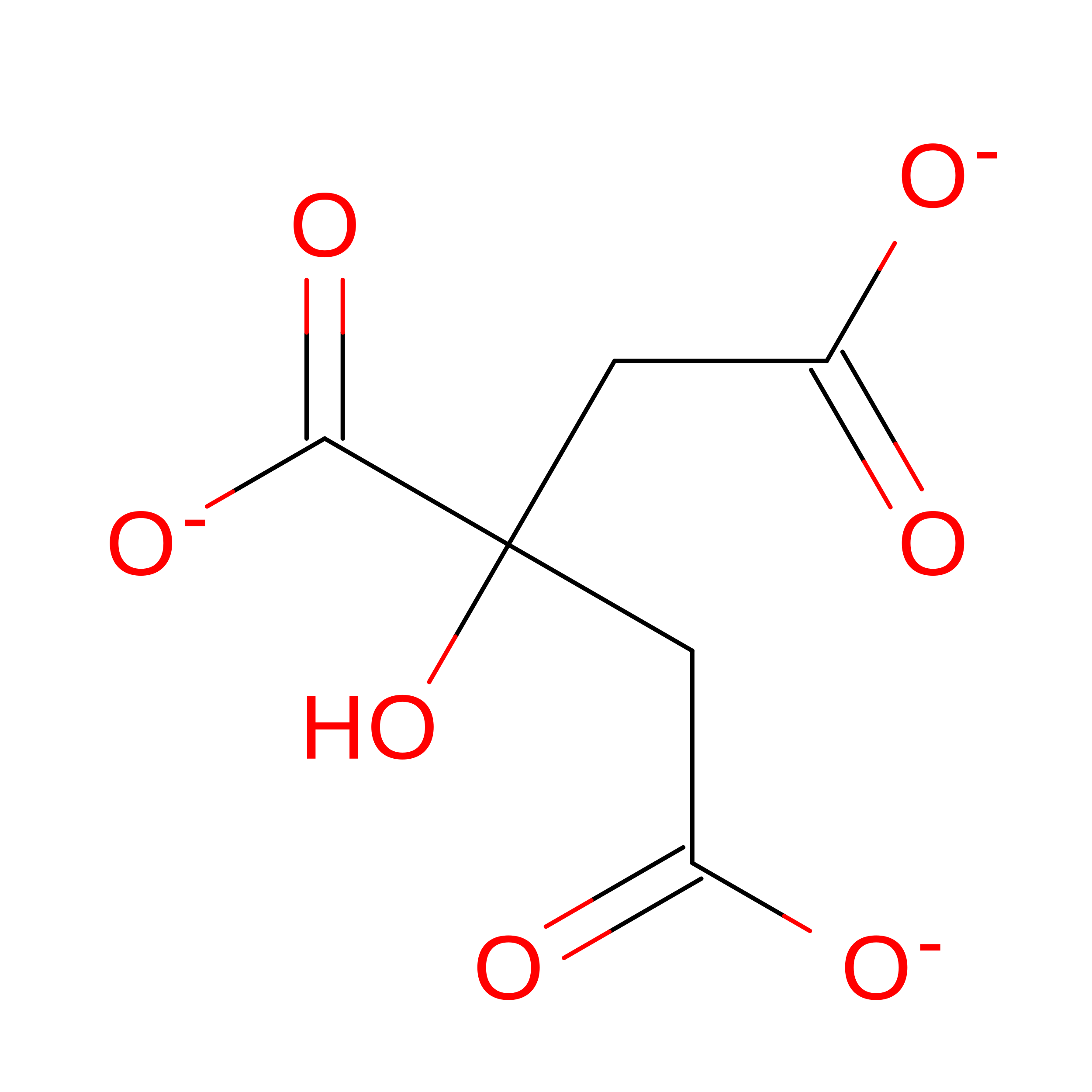
CITRATE ANION
C6 H5 O7
KRKNYBCHXYNGOX-UHFFFAOYSA-K
GOL

Query on GOL


Download:Ideal Coordinates CCD File
H [auth B],
K [auth C]
GLYCEROL
C3 H8 O3
PEDCQBHIVMGVHV-UHFFFAOYSA-N
CL

Query on CL


Download:Ideal Coordinates CCD File
I [auth B]CHLORIDE ION
Cl
VEXZGXHMUGYJMC-UHFFFAOYSA-M

Experimental Data & Validation

Experimental Data

  • Method: X-RAY DIFFRACTION
  • Resolution: 2.98 Å
  • R-Value Free:  0.259 (Depositor), 0.259 (DCC) 
  • R-Value Work:  0.212 (Depositor), 0.218 (DCC) 
  • R-Value Observed: 0.217 (Depositor) 
Space Group: P 65 2 2
Unit Cell:
Length ( Å )Angle ( ˚ )
a = 83.83α = 90
b = 83.83β = 90
c = 484.4γ = 120
Software Package:
Software NamePurpose
PHENIXrefinement
XSCALEdata scaling
XDSdata reduction
PHASERphasing

Structure Validation

View Full Validation Report


Ligand Structure Quality Assessment 


Entry History 

& Funding Information

Deposition Data


Funding OrganizationLocationGrant Number
National Institutes of Health/National Institute of General Medical Sciences (NIH/NIGMS)United StatesR35GM133482

Revision History  (Full details and data files)

  • Version 1.0: 2026-02-25
    Type: Initial release
  • Version 1.1: 2026-09-09
    Changes: Database references