9MT4 | pdb_00009mt4

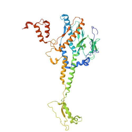
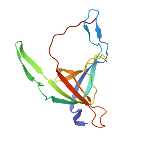
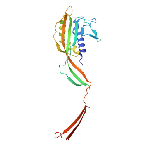
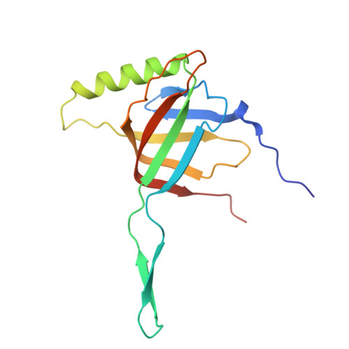
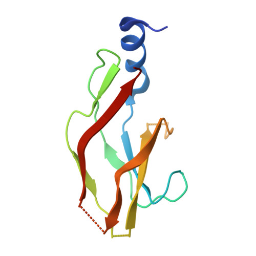
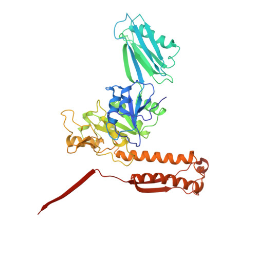
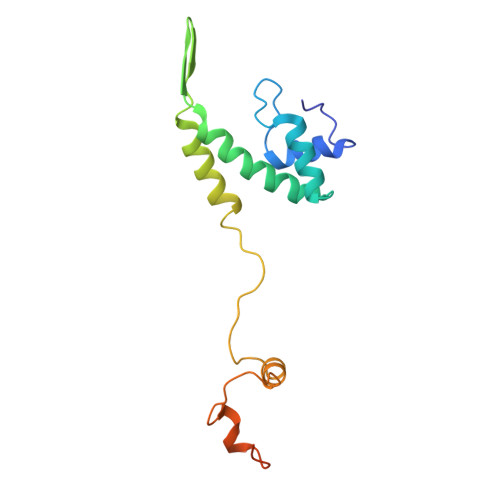
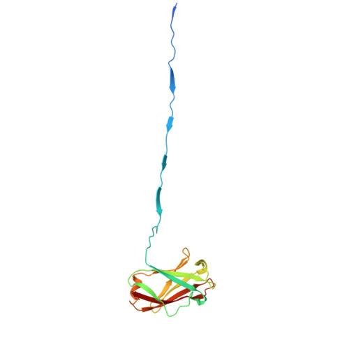
Connector structure of phage JohannRWettstein (Bas63)


Experimental Data Snapshot

  • Method: ELECTRON MICROSCOPY
  • Resolution: 3.10 Å
  • Aggregation State: PARTICLE 
  • Reconstruction Method: SINGLE PARTICLE 

wwPDB Validation 3D Report Full Report

Validation slider image for 9MT4

This is version 1.1 of the entry. See complete history

Literature

Cryo-EM structure of bacteriophage Bas63 reveals structural conservation and diversity in the Felixounavirus genus.

Hodgkinson-Bean, J.Ayala, R.McJarrow-Keller, K.Cassin, L.Rutter, G.L.Crowe, A.J.M.Wolf, M.Bostina, M.

(2025) Sci Adv 11: eadx0790-eadx0790

  • DOI: https://doi.org/10.1126/sciadv.adx0790
  • Primary Citation Related Structures: 
    9MT4, 9MT5

  • PubMed Abstract: 

    The BASEL phage collection was developed to provide access to diverse bacteriophages, distinct from model phages. Escherichia phage JohannRWettstein (Bas63), a myophage in the collection, is a member of the subfamily Ounavirinae and the Felixounavirus genus. Using cryo-electron microscopy, we investigated Bas63's structure to explore its evolutionary relationships and functional adaptations. Our structures reveal a series of gene products: (i) a capsid decorated with β-tulip proteins at three-fold symmetry axes and a Hoc-like protein at hexamer centers, (ii) a conserved connector with an additional 12-fold ring of collar proteins that extend unique whisker proteins that are structurally related to podophage GP4 tail fibers, and (iii) a baseplate with long tail fibers resembling a contracted form of T4's long tail fibers. Sequence conservation analysis of Bas63 structural proteins across ICTV-recognized Felixounavirus' supports its role as a structural model for Felixounavirus evolution. This study advances the mechanistic understanding of phage architecture and reinforces the structural mosaicism of bacteriophages.


  • Organizational Affiliation
    • Department of Microbiology and Immunology, University of Otago, Dunedin, New Zealand.

Macromolecule Content 

  • Total Structure Weight: 419.66 kDa 
  • Atom Count: 27,234 
  • Modeled Residue Count: 3,495 
  • Deposited Residue Count: 3,794 
  • Unique protein chains: 8

Macromolecules

Find similar proteins by:|  3D Structure
Entity ID: 1
MoleculeChains  Sequence LengthOrganismDetailsImage
Portal proteinA [auth B],
B [auth A]
488Escherichia phage JohannRWettsteinMutation(s): 0 
UniProt
Find proteins for A0AAE7VVG7 (Escherichia phage JohannRWettstein)
Explore A0AAE7VVG7 
Go to UniProtKB:  A0AAE7VVG7
Entity Groups
Sequence Clusters30% Identity50% Identity70% Identity90% Identity95% Identity100% Identity
UniProt GroupA0AAE7VVG7
Sequence Annotations
Expand
Reference Sequence
Find similar proteins by:|  3D Structure
Entity ID: 2
MoleculeChains  Sequence LengthOrganismDetailsImage
Head-closure proteinC [auth E]133Escherichia phage JohannRWettsteinMutation(s): 0 
UniProt
Find proteins for A0AAE7VV34 (Escherichia phage JohannRWettstein)
Explore A0AAE7VV34 
Go to UniProtKB:  A0AAE7VV34
Entity Groups
Sequence Clusters30% Identity50% Identity70% Identity90% Identity95% Identity100% Identity
UniProt GroupA0AAE7VV34
Sequence Annotations
Expand
Reference Sequence
Find similar proteins by:|  3D Structure
Entity ID: 3
MoleculeChains  Sequence LengthOrganismDetailsImage
Tail terminator proteinD [auth F]199Escherichia phage JohannRWettsteinMutation(s): 0 
UniProt
Find proteins for A0AAE7VUU6 (Escherichia phage JohannRWettstein)
Explore A0AAE7VUU6 
Go to UniProtKB:  A0AAE7VUU6
Entity Groups
Sequence Clusters30% Identity50% Identity70% Identity90% Identity95% Identity100% Identity
UniProt GroupA0AAE7VUU6
Sequence Annotations
Expand
Reference Sequence
Find similar proteins by:|  3D Structure
Entity ID: 4
MoleculeChains  Sequence LengthOrganismDetailsImage
Tube proteinE [auth P],
F [auth O]
148Escherichia phage JohannRWettsteinMutation(s): 0 
UniProt
Find proteins for A0AAE7VV62 (Escherichia phage JohannRWettstein)
Explore A0AAE7VV62 
Go to UniProtKB:  A0AAE7VV62
Entity Groups
Sequence Clusters30% Identity50% Identity70% Identity90% Identity95% Identity100% Identity
UniProt GroupA0AAE7VV62
Sequence Annotations
Expand
Reference Sequence
Find similar proteins by:|  3D Structure
Entity ID: 5
MoleculeChains  Sequence LengthOrganismDetailsImage
Phage protein110Escherichia phage JohannRWettsteinMutation(s): 0 
UniProt
Find proteins for A0AAE7VWD1 (Escherichia phage JohannRWettstein)
Explore A0AAE7VWD1 
Go to UniProtKB:  A0AAE7VWD1
Entity Groups
Sequence Clusters30% Identity50% Identity70% Identity90% Identity95% Identity100% Identity
UniProt GroupA0AAE7VWD1
Sequence Annotations
Expand
Reference Sequence
Find similar proteins by:|  3D Structure
Entity ID: 6
MoleculeChains  Sequence LengthOrganismDetailsImage
Structural proteinM [auth Q],
N [auth R]
450Escherichia phage JohannRWettsteinMutation(s): 0 
UniProt
Find proteins for A0AAE7VVP5 (Escherichia phage JohannRWettstein)
Explore A0AAE7VVP5 
Go to UniProtKB:  A0AAE7VVP5
Entity Groups
Sequence Clusters30% Identity50% Identity70% Identity90% Identity95% Identity100% Identity
UniProt GroupA0AAE7VVP5
Sequence Annotations
Expand
Reference Sequence
Find similar proteins by:|  3D Structure
Entity ID: 7
MoleculeChains  Sequence LengthOrganismDetailsImage
Phage proteinO [auth C],
P [auth D]
149Escherichia phage JohannRWettsteinMutation(s): 0 
UniProt
Find proteins for A0AAE8B2G2 (Escherichia phage JohannRWettstein)
Explore A0AAE8B2G2 
Go to UniProtKB:  A0AAE8B2G2
Entity Groups
Sequence Clusters30% Identity50% Identity70% Identity90% Identity95% Identity100% Identity
UniProt GroupA0AAE8B2G2
Sequence Annotations
Expand
Reference Sequence
Find similar proteins by:|  3D Structure
Entity ID: 8
MoleculeChains  Sequence LengthOrganismDetailsImage
Collar proteinQ [auth G],
R [auth H]
166Escherichia phage JohannRWettsteinMutation(s): 0 
UniProt
Find proteins for A0AAE7VUQ8 (Escherichia phage JohannRWettstein)
Explore A0AAE7VUQ8 
Go to UniProtKB:  A0AAE7VUQ8
Entity Groups
Sequence Clusters30% Identity50% Identity70% Identity90% Identity95% Identity100% Identity
UniProt GroupA0AAE7VUQ8
Sequence Annotations
Expand
Reference Sequence

Experimental Data & Validation

Experimental Data

  • Method: ELECTRON MICROSCOPY
  • Resolution: 3.10 Å
  • Aggregation State: PARTICLE 
  • Reconstruction Method: SINGLE PARTICLE 

Structure Validation

View Full Validation Report


Entry History 

& Funding Information

Deposition Data


Funding OrganizationLocationGrant Number
Ministry of Agriculture, Forestry and Fisheries (MAFF)JapanGD0696J004
Japan Society for the Promotion of Science (JSPS)Japan21K20645

Revision History  (Full details and data files)

  • Version 1.0: 2025-11-19
    Type: Initial release
  • Version 1.1: 2025-11-26
    Changes: Data collection, Database references