9K3N | pdb_00009k3n

The structure of Salmonella phage PJNS002


Experimental Data Snapshot

  • Method: ELECTRON MICROSCOPY
  • Resolution: 2.59 Å
  • Aggregation State: PARTICLE 
  • Reconstruction Method: SINGLE PARTICLE 

Starting Model: in silico
View more details

wwPDB Validation 3D Report Full Report

Validation slider image for 9K3N

This is version 1.1 of the entry. See complete history

Literature

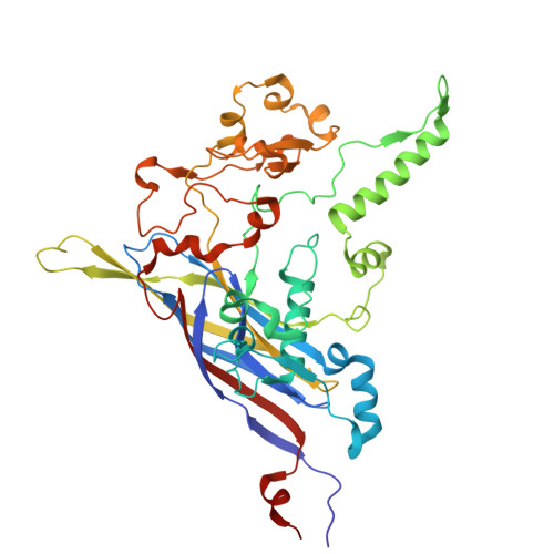
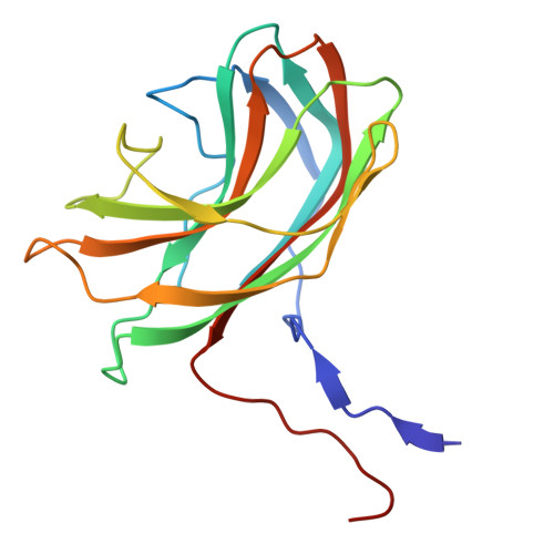
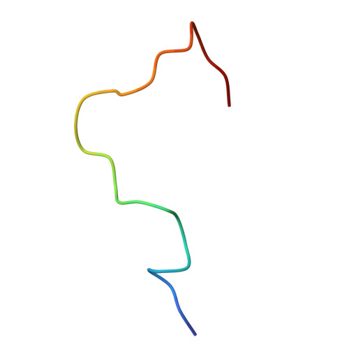
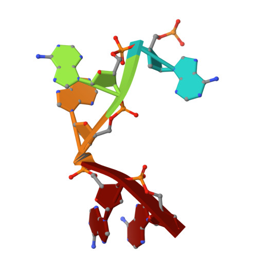
Structural basis for Salmonella infection by two Microviridae phages.

Hu, W.Liu, Z.Wei, Y.Bian, Q.Lan, W.Fan, C.Song, J.Sun, Q.Zhang, X.Liu, Y.Gao, Y.Chen, Y.

(2025) Commun Biol 8: 1166-1166

  • DOI: https://doi.org/10.1038/s42003-025-08595-7
  • Primary Citation Related Structures: 
    9K3M, 9K3N

  • PubMed Abstract: 

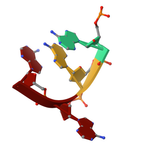
    The global resurgence of multidrug-resistant Salmonella species, responsible for millions of annual infections, underscores the urgent need for alternative antimicrobial strategies, such as phage therapy. Microviridae phages offer a promising model for studying phage-host interactions with their unique structural and infection mechanisms. Here, we identify two Microviridae phages, PJNS001 and PJNS002, with different host receptor dependencies, and determine their cryo-EM structures at 2.68 Å and 2.59 Å resolution, respectively. These icosahedral capsids with T = 1 symmetry exhibit a unique vertex reinforcement mechanism, stabilizing the viral assembly. The specific pentameric adaptations, coupled with DNA binding protein engagements and thermodynamic constraints, collectively preclude the formation of hybrid virions. Structural analysis and in situ visualization reveal spike protein features and host-attachment intermediates, informing host specificity. Together, these findings advance our understanding of Microviridae infection mechanisms and provide a structural framework for rational phage design against antibiotic-resistant pathogens.


  • Organizational Affiliation
    • Shanghai Institute for Advanced Immunochemical Studies, ShanghaiTech University, Shanghai, 201210, China.

Macromolecule Content 

  • Total Structure Weight: 4,376.47 kDa 
  • Atom Count: 307,380 
  • Modeled Residue Count: 38,400 
  • Deposited Residue Count: 38,460 
  • Unique protein chains: 3
  • Unique nucleic acid chains: 2

Macromolecules


Find similar proteins by:|  3D Structure
Find similar nucleic acids by:  Sequence
Entity ID: 4
MoleculeChains LengthOrganismImage
DNA (5'-D(P*AP*AP*AP*AP*A)-3')5Salmonella phage PJNS002
Sequence Annotations
Expand
Reference Sequence
Find similar nucleic acids by:  Sequence
Entity ID: 5
MoleculeChains LengthOrganismImage
DNA (5'-D(P*AP*AP*AP*A)-3')4Salmonella phage PJNS002
Sequence Annotations
Expand
Reference Sequence

Experimental Data & Validation

Experimental Data

  • Method: ELECTRON MICROSCOPY
  • Resolution: 2.59 Å
  • Aggregation State: PARTICLE 
  • Reconstruction Method: SINGLE PARTICLE 
EM Software:
TaskSoftware PackageVersion
RECONSTRUCTIONcryoSPARC4.0.1
MODEL REFINEMENTPHENIX1.20.1

Structure Validation

View Full Validation Report


Entry History 

& Funding Information

Deposition Data


Funding OrganizationLocationGrant Number
Other governmentChina23QA1406400

Revision History  (Full details and data files)

  • Version 1.0: 2025-08-13
    Type: Initial release
  • Version 1.1: 2025-08-20
    Changes: Data collection, Database references