9K0K | pdb_00009k0k

Cryo-EM structure of UTP-bound P2Y purinoceptor 4-miniGq-Nb35 complex


Experimental Data Snapshot

  • Method: ELECTRON MICROSCOPY
  • Resolution: 3.14 Å
  • Aggregation State: PARTICLE 
  • Reconstruction Method: SINGLE PARTICLE 

wwPDB Validation 3D Report Full Report

Validation slider image for 9K0K

This is version 1.1 of the entry. See complete history

Literature

Structural insight into the self-activation and G-protein coupling of P2Y2 receptor.

Lan, B.Zhang, S.Chen, K.Dai, S.Fei, J.Gao, K.Sun, X.Lin, B.Liu, X.

(2025) Cell Discov 11: 47-47

  • DOI: https://doi.org/10.1038/s41421-025-00797-x
  • Primary Citation Related Structures: 
    9K0K, 9K0X, 9K20, 9K25

  • PubMed Abstract: 

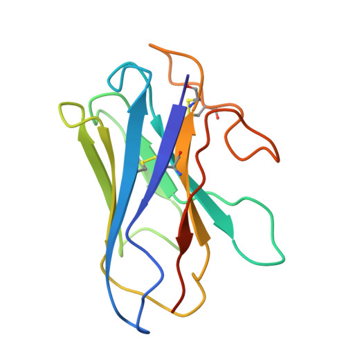
    Purinergic P2Y2 receptor (P2Y2R) represents a typically extracellular ATP and UTP sensor for mediating purinergic signaling. Despite its importance as a pharmacological target, the molecular mechanisms underlying ligand recognition and G-protein coupling have remained elusive due to lack of structural information. In this study, we determined the cryo-electron microscopy (cryo-EM) structures of the apo P2Y2R in complex with G q , ATP-bound P2Y2R in complex with G q or G o , and UTP-bound P2Y4R in complex with G q . These structures reveal the similarities and distinctions of ligand recognition within the P2Y receptor family. Furthermore, a comprehensive analysis of G-protein coupling reveals that P2Y2R exhibits promiscuity in coupling with both G q and G o proteins. Combining molecular dynamics simulations and signaling assays, we elucidate the molecular mechanisms by which P2Y2R differentiates pathway-specific G q or G o coupling through distinct structural components on the intracellular side. Strikingly, we identify a helix-like segment within the N-terminus that occupies the orthosteric ligand-binding pocket of P2Y2R, accounting for its self-activation. Taken together, these findings provide a molecular framework for understanding the activation mechanism of P2Y2R, encompassing ligand recognition, G-protein coupling, and a novel N-terminus-mediated self-activation mechanism.


  • Organizational Affiliation
    • State Key Laboratory of Membrane Biology, Tsinghua-Peking Center for Life Sciences, School of Pharmaceutical Sciences, Tsinghua University, Beijing, China.

Macromolecule Content 

  • Total Structure Weight: 136.25 kDa 
  • Atom Count: 7,441 
  • Modeled Residue Count: 996 
  • Deposited Residue Count: 1,214 
  • Unique protein chains: 5

Macromolecules

Find similar proteins by:|  3D Structure
Entity ID: 1
MoleculeChains  Sequence LengthOrganismDetailsImage
Guanine nucleotide-binding protein G(s) subunit alpha isoforms short243Homo sapiensMutation(s): 0 
Gene Names: GNASGNAS1GSP
EC: 3.6.5
Membrane Entity: Yes 
UniProt & NIH Common Fund Data Resources
Find proteins for P63092 (Homo sapiens)
Explore P63092 
Go to UniProtKB:  P63092
PHAROS:  P63092
GTEx:  ENSG00000087460 
Entity Groups
Sequence Clusters30% Identity50% Identity70% Identity90% Identity95% Identity100% Identity
UniProt GroupP63092
Sequence Annotations
Expand
Reference Sequence
Find similar proteins by:|  3D Structure
Entity ID: 2
MoleculeChains  Sequence LengthOrganismDetailsImage
Guanine nucleotide-binding protein G(I)/G(S)/G(T) subunit beta-1358Homo sapiensMutation(s): 0 
Gene Names: GNB1
UniProt & NIH Common Fund Data Resources
Find proteins for P62873 (Homo sapiens)
Explore P62873 
Go to UniProtKB:  P62873
PHAROS:  P62873
GTEx:  ENSG00000078369 
Entity Groups
Sequence Clusters30% Identity50% Identity70% Identity90% Identity95% Identity100% Identity
UniProt GroupP62873
Sequence Annotations
Expand
Reference Sequence
Find similar proteins by:|  3D Structure
Entity ID: 3
MoleculeChains  Sequence LengthOrganismDetailsImage
Guanine nucleotide-binding protein G(I)/G(S)/G(O) subunit gamma-2C [auth G]71Homo sapiensMutation(s): 0 
Gene Names: GNG2
Membrane Entity: Yes 
UniProt & NIH Common Fund Data Resources
Find proteins for P59768 (Homo sapiens)
Explore P59768 
Go to UniProtKB:  P59768
PHAROS:  P59768
GTEx:  ENSG00000186469 
Entity Groups
Sequence Clusters30% Identity50% Identity70% Identity90% Identity95% Identity100% Identity
UniProt GroupP59768
Sequence Annotations
Expand
Reference Sequence
Find similar proteins by:|  3D Structure
Entity ID: 4
MoleculeChains  Sequence LengthOrganismDetailsImage
Nanobody 35D [auth N]134Vicugna pacosMutation(s): 0 
Find similar proteins by:|  3D Structure
Entity ID: 5
MoleculeChains  Sequence LengthOrganismDetailsImage
P2Y purinoceptor 4E [auth I]408Homo sapiensMutation(s): 0 
Gene Names: P2RY4NRU
Membrane Entity: Yes 
UniProt & NIH Common Fund Data Resources
Find proteins for P51582 (Homo sapiens)
Explore P51582 
Go to UniProtKB:  P51582
PHAROS:  P51582
GTEx:  ENSG00000186912 
Entity Groups
Sequence Clusters30% Identity50% Identity70% Identity90% Identity95% Identity100% Identity
UniProt GroupP51582
Sequence Annotations
Expand
Reference Sequence

Small Molecules

Ligands 1 Unique
IDChains Name / Formula / InChI Key2D Diagram3D Interactions
UTP
(Subject of Investigation/LOI)

Query on UTP


Download:Ideal Coordinates CCD File
F [auth I]URIDINE 5'-TRIPHOSPHATE
C9 H15 N2 O15 P3
PGAVKCOVUIYSFO-XVFCMESISA-N

Experimental Data & Validation

Experimental Data

  • Method: ELECTRON MICROSCOPY
  • Resolution: 3.14 Å
  • Aggregation State: PARTICLE 
  • Reconstruction Method: SINGLE PARTICLE 
EM Software:
TaskSoftware PackageVersion
MODEL REFINEMENTPHENIX

Structure Validation

View Full Validation Report


Entry History 

& Funding Information

Deposition Data


Funding OrganizationLocationGrant Number
National Natural Science Foundation of China (NSFC)China--

Revision History  (Full details and data files)

  • Version 1.0: 2025-06-11
    Type: Initial release
  • Version 1.1: 2025-07-23
    Changes: Data collection