Integrative Approach to Develop and Characterize Antibodies against the Cancer-Associated Antigen Sialyl Lewis A (CA 19-9).
Freitag, A., Khilji, S.K., Nedielkov, R., M Kumar, S., Krummhaar, M., Arndt, J., Moreira, G.M.S.G., Luhle, J., Goerdeler, F., Kamphues, C., Mroginski, M.A., Roth, C., Seeberger, P.H., Moller, H.M., Moscovitz, O.(2026) JACS Au 6: 2274-2287
- PubMed: 42063835 Search on PubMedSearch on PubMed Central
- DOI: https://doi.org/10.1021/jacsau.5c01596
- Primary Citation Related Structures:
9I6Q, 9I9H - PubMed Abstract:
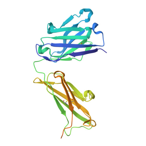
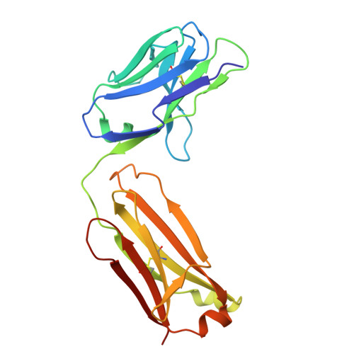
Sialyl Lewis A (sLeA), or the CA 19-9 marker, is the best validated and only FDA-approved serologic marker clinically used to monitor recurrence, progression, and therapy efficiency in pancreatic ductal adenocarcinoma (PDAC) patients, making it an attractive target for antibody development. Recent clinical trials have demonstrated satisfying safety profiles and unique expression in a range of malignancies that highlight CA 19-9 as an attractive TACA to target using a stand-alone drug or an adjuvant therapy. Hence, we set out to explore the use of synthetic sLeA in the development of additional monoclonal antibodies (mAbs) with enhanced sLeA recognition and improved efficacy. Two mAbs targeting sLeA were generated through mice immunization with synthetic sLeA glycoconjugates, synthetic glycan arrays, and hybridoma technology. We then compared the antigen-binding properties of the newly developed mAbs with those of the widely used mAb 1116-NS-19-9 and demonstrated improved affinity and specificity for native sLeA ectopically expressed in B16 melanoma cells, surpassing the performance of the established mAb 1116-NS-19-9. Of the two mAbs, GB11 was more promising. Therefore, to elucidate the structural origin of improved GB11's antigen binding, we conducted high-resolution mapping of the molecular recognition patterns between sLeA and the different antibodies using X-ray crystallography and STD NMR. These analyses revealed subtle yet critical differences in the glycan engagement and identified key structural features underlying enhanced GB11's recognition of sLeA. MD simulations further supported these observations, indicating distinct orientations of sLeA within the binding pockets of each mAb. Our results suggest improved recognition of the native sLeA antigen by the newly generated GB11 antibody, providing a detailed and high-resolution elucidation of the molecular interactions underlying this recognition. Our study provides a tool with improved theranostic properties against sLeA-overexpressing malignancies.
- Department of Biomolecular Systems, Max Planck Institute of Colloids and Interfaces, 14476 Potsdam, Germany.
Organizational Affiliation: