In situ and in vitro cryo-EM reveal structures of mycobacterial encapsulin assembly intermediates.
Berger, C., Lewis, C., Gao, Y., Knoops, K., Lopez-Iglesias, C., Peters, P.J., Ravelli, R.B.G.(2025) Commun Biol 8: 245-245
- PubMed: 39955411 Search on PubMedSearch on PubMed Central
- DOI: https://doi.org/10.1038/s42003-025-07660-5
- Primary Citation Related Structures:
7P1T, 9GOT, 9HQ7, 9HQC - PubMed Abstract:
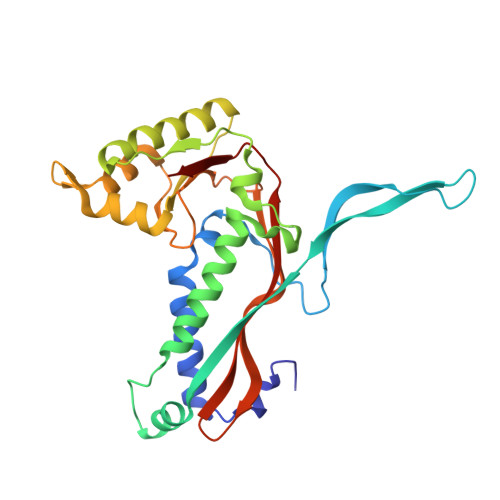
Prokaryotes rely on proteinaceous compartments such as encapsulin to isolate harmful reactions. Encapsulin are widely expressed by bacteria, including the Mycobacteriaceae, which include the human pathogens Mycobacterium tuberculosis and Mycobacterium leprae. Structures of fully assembled encapsulin shells have been determined for several species, but encapsulin assembly and cargo encapsulation are still poorly characterised, because of the absence of encapsulin structures in intermediate assembly states. We combine in situ and in vitro structural electron microscopy to show that encapsulins are dynamic assemblies with intermediate states of cargo encapsulation and shell assembly. Using cryo-focused ion beam (FIB) lamella preparation and cryo-electron tomography (CET), we directly visualise encapsulins in Mycobacterium marinum, and observed ribbon-like attachments to the shell, encapsulin shells with and without cargoes, and encapsulin shells in partially assembled states. In vitro cryo-electron microscopy (EM) single-particle analysis of the Mycobacterium tuberculosis encapsulin was used to obtain three structures of the encapsulin shell in intermediate states, as well as a 2.3 Å structure of the fully assembled shell. Based on the analysis of the intermediate encapsulin shell structures, we propose a model of encapsulin self-assembly via the pairwise addition of monomers.
- Division of Nanoscopy, Maastricht Multimodal Molecular Imaging Institute, Maastricht University, Maastricht, The Netherlands. casper.berger@rfi.ac.uk.
Organizational Affiliation: