9GM2 | pdb_00009gm2

Human SMUG1 in complex with DNA


Experimental Data Snapshot

  • Method: X-RAY DIFFRACTION
  • Resolution: 2.10 Å
  • R-Value Free: 
    0.253 (Depositor), 0.253 (DCC) 
  • R-Value Work: 
    0.207 (Depositor), 0.206 (DCC) 

Starting Model: experimental
View more details

wwPDB Validation 3D Report Full Report

Validation slider image for 9GM2

This is version 1.1 of the entry. See complete history

Literature

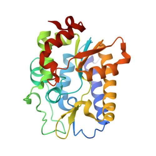
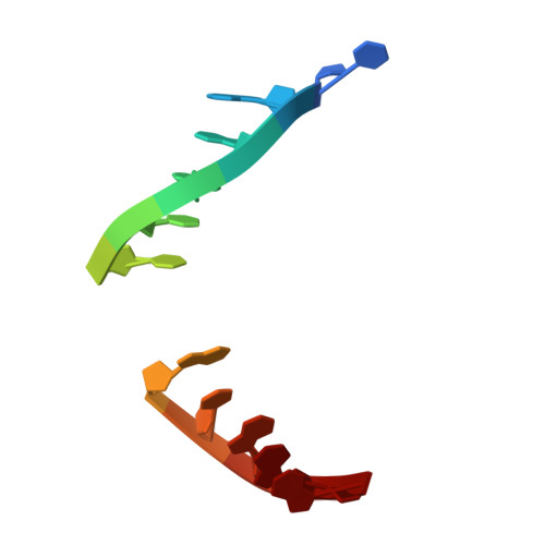
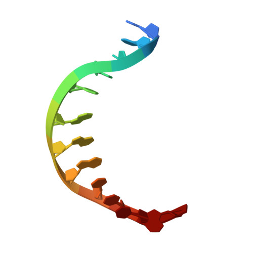
Structural basis for uracil removal from DNA by human SMUG1.

Ludascher, J.M.Scaletti Hutchinson, E.Vila-Julia, G.Jemth, A.S.Shahid, S.Wiita, E.Cabeza de Vaca, I.Pach, S.Gajdos, L.Aggarwal, S.Walse, E.Mortusewicz, O.Helleday, T.Carlsson, J.Stenmark, P.

(2026) Nat Commun 17

  • DOI: https://doi.org/10.1038/s41467-026-72937-0
  • Primary Citation Related Structures: 
    9GGS, 9GK0, 9GM2, 9RQP, 9RQS

  • PubMed Abstract: 

    Human single-strand-selective monofunctional uracil DNA glycosylase 1 (hSMUG1) removes uracil, 5-hydroxymethyluracil (5hmU) and 5-fluorouracil (5FU) from DNA, thereby initiating the base excision repair (BER) process. hSMUG1 is important for maintaining genomic integrity and plays a significant role in cancer biology. Here, we present the structures of hSMUG1, including complexes with products (uracil and 5FU) and an enzyme-product complex of hSMUG1 with double-stranded DNA (dsDNA). Analysis of our hSMUG1-dsDNA complex reveals how uracil is flipped out of the dsDNA for excision and identifies key residues that we confirm to be critical for both DNA binding and enzymatic activity. Furthermore, our hSMUG1 substrate complexes, molecular dynamics simulations and neutron diffraction data suggest a mechanism by which the substrate uracil rotates following base excision. The structural and functional information presented here will be highly useful for the future development of inhibitors and/or activators targeting hSMUG1.


  • Organizational Affiliation
    • Department of Biochemistry and Biophysics, Stockholm University, Stockholm, Sweden.

Macromolecule Content 

  • Total Structure Weight: 124.31 kDa 
  • Atom Count: 9,014 
  • Modeled Residue Count: 1,036 
  • Deposited Residue Count: 1,036 
  • Unique protein chains: 1
  • Unique nucleic acid chains: 2

Macromolecules


Find similar proteins by:|  3D Structure
Entity ID: 1
MoleculeChains  Sequence LengthOrganismDetailsImage
Single-strand selective monofunctional uracil DNA glycosylase
A, B, C, D
247Homo sapiensMutation(s): 0 
Gene Names: SMUG1
EC: 3.2.2
UniProt & NIH Common Fund Data Resources
Find proteins for Q53HV7 (Homo sapiens)
Explore Q53HV7 
Go to UniProtKB:  Q53HV7
PHAROS:  Q53HV7
GTEx:  ENSG00000123415 
Entity Groups
Sequence Clusters30% Identity50% Identity70% Identity90% Identity95% Identity100% Identity
UniProt GroupQ53HV7
Sequence Annotations
Expand
Reference Sequence
Find similar nucleic acids by:  Sequence
Entity ID: 2
MoleculeChains LengthOrganismImage
DNA (5'-D(P*CP*GP*GP*AP*CP*T*ORP*AP*CP*GP*GP*G)-3')
E, G
12Homo sapiens
Sequence Annotations
Expand
Reference Sequence
Find similar nucleic acids by:  Sequence
Entity ID: 3
MoleculeChains LengthOrganismImage
DNA (5'-D(P*CP*CP*CP*GP*TP*GP*AP*GP*TP*CP*CP*G)-3')
F, H
12Homo sapiens
Sequence Annotations
Expand
Reference Sequence

Experimental Data & Validation

Experimental Data

  • Method: X-RAY DIFFRACTION
  • Resolution: 2.10 Å
  • R-Value Free:  0.253 (Depositor), 0.253 (DCC) 
  • R-Value Work:  0.207 (Depositor), 0.206 (DCC) 
Space Group: C 1 2 1
Unit Cell:
Length ( Å )Angle ( ˚ )
a = 150.072α = 90
b = 84.366β = 113.403
c = 88.259γ = 90
Software Package:
Software NamePurpose
REFMACrefinement
XDSdata reduction
STARANISOdata scaling
PHASERphasing

Structure Validation

View Full Validation Report


Entry History 

& Funding Information

Deposition Data


Funding OrganizationLocationGrant Number
CancerfondenSweden--

Revision History  (Full details and data files)

  • Version 1.0: 2026-03-11
    Type: Initial release
  • Version 1.1: 2026-06-17
    Changes: Database references