


Crystal structure of human Haspin (GSG2) kinase bound to MU1959
Kraemer, A., Paruch, K., Knapp, S., Structural Genomics Consortium (SGC)To be published.
Experimental Data Snapshot
Entity ID: 1 | |||||
|---|---|---|---|---|---|
| Molecule | Chains | Sequence Length | Organism | Details | Image |
| Serine/threonine-protein kinase haspin | 357 | Homo sapiens | Mutation(s): 0 Gene Names: HASPIN, GSG2 EC: 2.7.11.1 | 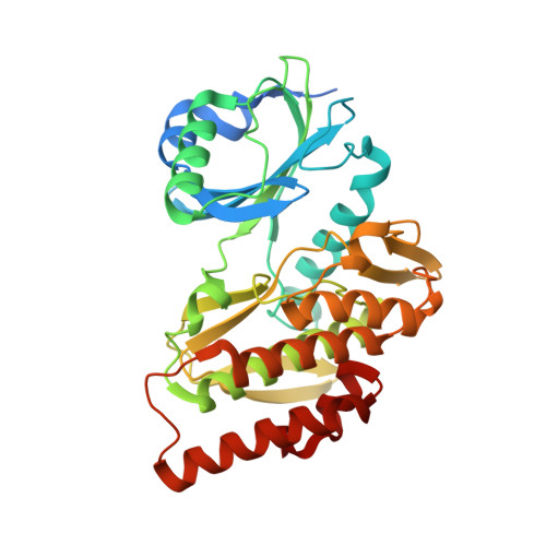 | |
UniProt & NIH Common Fund Data Resources | |||||
PHAROS: Q8TF76 GTEx: ENSG00000177602 | |||||
Entity Groups | |||||
| Sequence Clusters | 30% Identity50% Identity70% Identity90% Identity95% Identity100% Identity | ||||
| UniProt Group | Q8TF76 | ||||
Sequence AnnotationsExpand | |||||
Reference Sequence | |||||
| Ligands 5 Unique | |||||
|---|---|---|---|---|---|
| ID | Chains | Name / Formula / InChI Key | 2D Diagram | 3D Interactions | |
| A1IDD (Subject of Investigation/LOI) Download:Ideal Coordinates CCD File | G [auth A] | [4-[5-(1-methylpyrazol-4-yl)pyrazolo[1,5-a]pyrimidin-3-yl]pyridin-2-yl]methanamine C16 H15 N7 FVQBDHAVXPEVEZ-UHFFFAOYSA-N |  | ||
| EPE Download:Ideal Coordinates CCD File | B [auth A] | 4-(2-HYDROXYETHYL)-1-PIPERAZINE ETHANESULFONIC ACID C8 H18 N2 O4 S JKMHFZQWWAIEOD-UHFFFAOYSA-N |  | ||
| MPD Download:Ideal Coordinates CCD File | C [auth A], F [auth A], H [auth A], I [auth A], J [auth A] | (4S)-2-METHYL-2,4-PENTANEDIOL C6 H14 O2 SVTBMSDMJJWYQN-YFKPBYRVSA-N |  | ||
| NI Download:Ideal Coordinates CCD File | K [auth A] | NICKEL (II) ION Ni VEQPNABPJHWNSG-UHFFFAOYSA-N |  | ||
| NA Download:Ideal Coordinates CCD File | D [auth A], E [auth A] | SODIUM ION Na FKNQFGJONOIPTF-UHFFFAOYSA-N |  | ||
| Length ( Å ) | Angle ( ˚ ) |
|---|---|
| a = 68.59 | α = 90 |
| b = 77.993 | β = 90 |
| c = 87.098 | γ = 90 |
| Software Name | Purpose |
|---|---|
| REFMAC | refinement |
| Aimless | data scaling |
| XDS | data reduction |
| MOLREP | phasing |
| Funding Organization | Location | Grant Number |
|---|---|---|
| Innovative Medicines Initiative | Switzerland | 875510 |


