9EQJ

Crystal structure of pVHL:EloB:EloC in complex with MP-1-39


Experimental Data Snapshot

  • Method: X-RAY DIFFRACTION
  • Resolution: 2.05 Å
  • R-Value Free: 0.216 
  • R-Value Work: 0.183 
  • R-Value Observed: 0.185 

Starting Model: experimental
View more details

wwPDB Validation   3D Report Full Report


Ligand Structure Quality Assessment 


This is version 1.0 of the entry. See complete history


Literature

Stereochemical inversion at a 1,4-cyclohexyl PROTAC linker fine-tunes conformation and binding affinity.

Pierri, M.Liu, X.Kroupova, A.Rutter, Z.Hallatt, A.J.Ciulli, A.

(2024) Bioorg Med Chem Lett 110: 129861-129861

  • DOI: https://doi.org/10.1016/j.bmcl.2024.129861
  • Primary Citation of Related Structures:  
    9EQJ, 9EQM

  • PubMed Abstract: 

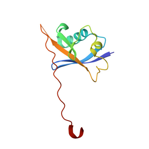
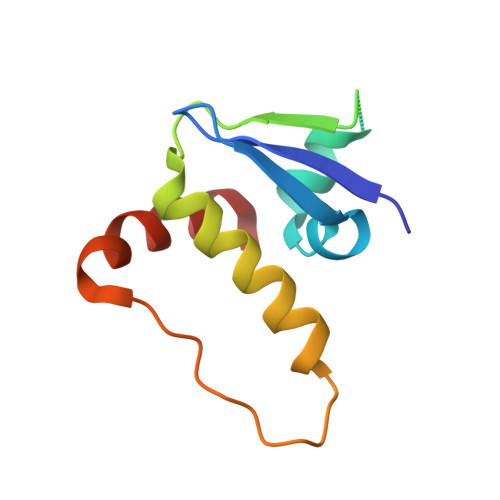
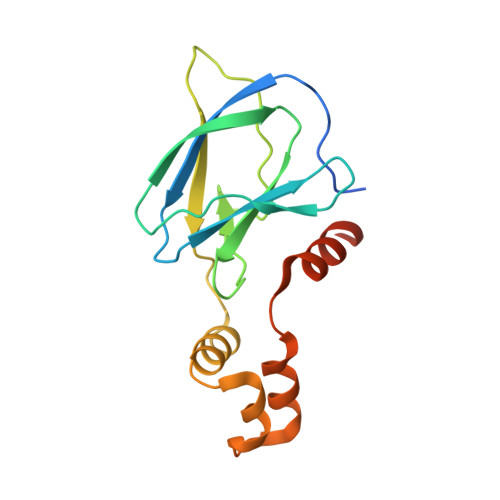
    Proteolysis targeting chimeras (PROTACs) are heterobifunctional small-molecule degraders made of a linker connecting a target-binding moiety to a ubiquitin E3 ligase-binding moiety. The linker unit is known to influence the physicochemical and pharmacokinetic properties of PROTACs, as well as the properties of ternary complexes, in turn impacting on their degradation activity in cells and in vivo. Our LRRK2 PROTAC XL01126, bearing a trans-cyclohexyl group in the linker, is a better and more cooperative degrader than its corresponding cis- analogue despite its much weaker binary binding affinities. Here, we investigate how this subtle stereocenter alteration in the linker affects the ligand binding affinity to the E3 ligase VHL. We designed a series of molecular matched pairs, truncating from the full PROTACs down to the VHL ligand, and find that across the series the trans-cyclohexyl compounds showed consistently weaker VHL-binding affinity compared to the cis- counterparts. High-resolution co-crystal structures revealed that the trans linker exhibits a rigid stick-out conformation, while the cis linker collapses into a folded-back conformation featuring a network of intramolecular contacts and long-range interactions with VHL. These observations are noteworthy as they reveal how a single stereochemical inversion within a PROTAC linker impacts conformational rigidity and binding mode, in turn fine-tuning differentiated propensity to binary and ternary complex formation, and ultimately cellular degradation activity.


  • Organizational Affiliation

    Centre for Targeted Protein Degradation, School of Life Sciences, University of Dundee, 1 James Lindsay Place, DD1 5JJ Dundee, United Kingdom.


Macromolecules
Find similar proteins by:  (by identity cutoff)  |  3D Structure
Entity ID: 1
MoleculeChains Sequence LengthOrganismDetailsImage
Elongin-B
A, D
104Homo sapiensMutation(s): 0 
Gene Names: ELOBTCEB2
UniProt & NIH Common Fund Data Resources
Find proteins for Q15370 (Homo sapiens)
Explore Q15370 
Go to UniProtKB:  Q15370
PHAROS:  Q15370
GTEx:  ENSG00000103363 
Entity Groups  
Sequence Clusters30% Identity50% Identity70% Identity90% Identity95% Identity100% Identity
UniProt GroupQ15370
Sequence Annotations
Expand
  • Reference Sequence
Find similar proteins by:  (by identity cutoff)  |  3D Structure
Entity ID: 2
MoleculeChains Sequence LengthOrganismDetailsImage
Elongin-C
B, E
97Homo sapiensMutation(s): 0 
Gene Names: ELOCTCEB1
UniProt & NIH Common Fund Data Resources
Find proteins for Q15369 (Homo sapiens)
Explore Q15369 
Go to UniProtKB:  Q15369
PHAROS:  Q15369
GTEx:  ENSG00000154582 
Entity Groups  
Sequence Clusters30% Identity50% Identity70% Identity90% Identity95% Identity100% Identity
UniProt GroupQ15369
Sequence Annotations
Expand
  • Reference Sequence
Find similar proteins by:  (by identity cutoff)  |  3D Structure
Entity ID: 3
MoleculeChains Sequence LengthOrganismDetailsImage
von Hippel-Lindau disease tumor suppressor
C, F
162Homo sapiensMutation(s): 0 
Gene Names: VHL
UniProt & NIH Common Fund Data Resources
Find proteins for P40337 (Homo sapiens)
Explore P40337 
Go to UniProtKB:  P40337
PHAROS:  P40337
GTEx:  ENSG00000134086 
Entity Groups  
Sequence Clusters30% Identity50% Identity70% Identity90% Identity95% Identity100% Identity
UniProt GroupP40337
Sequence Annotations
Expand
  • Reference Sequence
Small Molecules
Ligands 1 Unique
IDChains Name / Formula / InChI Key2D Diagram3D Interactions
A1H6O (Subject of Investigation/LOI)
Query on A1H6O

Download Ideal Coordinates CCD File 
G [auth C],
H [auth F]
(2S,4R)-1-[(2R)-2-[(1-fluoranylcyclopropyl)carbonylamino]-3-methyl-3-[[cis-4-(morpholin-4-ylmethyl)cyclohexyl]methylsulfanyl]butanoyl]-N-[[4-(4-methyl-1,3-thiazol-5-yl)phenyl]methyl]-4-oxidanyl-pyrrolidine-2-carboxamide
C37 H52 F N5 O5 S2
CLXVPFKDBLSVPK-WRZLIUBLSA-N
Experimental Data & Validation

Experimental Data

  • Method: X-RAY DIFFRACTION
  • Resolution: 2.05 Å
  • R-Value Free: 0.216 
  • R-Value Work: 0.183 
  • R-Value Observed: 0.185 
  • Space Group: P 1
Unit Cell:
Length ( Å )Angle ( ˚ )
a = 47.237α = 81.41
b = 47.345β = 76.78
c = 99.746γ = 80.49
Software Package:
Software NamePurpose
PHENIXrefinement
DIALSdata reduction
DIALSdata scaling
PHASERphasing

Structure Validation

View Full Validation Report


Ligand Structure Quality Assessment 


Entry History & Funding Information

Deposition Data


Funding OrganizationLocationGrant Number
Innovative Medicines InitiativeSwitzerland875510
UK Research and Innovation (UKRI)United KingdomEP/X025225/1

Revision History  (Full details and data files)

  • Version 1.0: 2024-08-28
    Type: Initial release