9CA9 | pdb_00009ca9

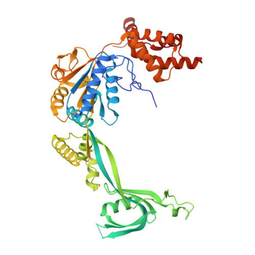
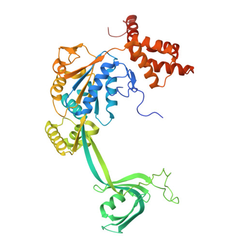
Cryo-EM structure of the human SRCAP complex in the unbound state (composite structure)

  • Classification: GENE REGULATION
  • Organism(s): Homo sapiens
  • Mutation(s): No 

  • Deposited: 2024-06-17 Released: 2025-06-18 
  • Deposition Author(s): Louder, R.K., Park, G.
  • Funding Organization(s): National Institutes of Health/National Institute of General Medical Sciences (NIH/NIGMS)

Experimental Data Snapshot

  • Method: ELECTRON MICROSCOPY
  • Resolution: 3.56 Å
  • Aggregation State: PARTICLE 
  • Reconstruction Method: SINGLE PARTICLE 

wwPDB Validation 3D Report Full Report

Validation slider image for 9CA9

This is version 1.1 of the entry. See complete history

Literature

Structural mechanism of histone H2A.Z exchange by human SRCAP-CFDP1 holoenzyme.

Park, G.Wu, C.Louder, R.K.

(2026) Sci Adv 12: eaei7728-eaei7728

  • DOI: https://doi.org/10.1126/sciadv.aei7728
  • Primary Citation Related Structures: 
    9CA7, 9CA8, 9CA9, 9CAA, 9NFV, 9NFW, 9NFX, 9NFY, 9NFZ, 9NG0, 9PGC, 9PGD, 9Y3D, 9Y3E, 9Y3F, 9Y3G, 9Y3H, 9ZLA

  • PubMed Abstract: 

    The conserved yeast SWR1 and human SRCAP chromatin remodeling complexes catalyze exchange of nucleosomal histone H2A for H2A.Z, but the underlying mechanism has remained obscure. Here, we show that histone exchange by SRCAP requires the transient activator CFDP1 and resolve nine cryo-electron microscopy structures of the SRCAP-CFDP1 holoenzyme that define the stepwise exchange mechanism. CFDP1 recognizes the conformation of the fully engaged SRCAP-nucleosome complex through interactions with multiple subunits-including direct contact with the ATPase domain-and induces conformational transitions that drive extensive DNA unwrapping, eviction of the H2A-H2B dimer, and insertion of the H2A.Z-H2B dimer, all without necessarily requiring hydrolysis of bound ATP. Collectively, these findings provide unprecedented insight into the mechanism of activator- and nucleotide-driven histone exchange from nucleosomal H2A to H2A.Z.


  • Organizational Affiliation
    • Biochemistry, Cellular and Molecular Biology Graduate Program, Johns Hopkins University School of Medicine, Baltimore, MD 21205, USA.

Macromolecule Content 

  • Total Structure Weight: 755.87 kDa 
  • Atom Count: 28,589 
  • Modeled Residue Count: 3,618 
  • Deposited Residue Count: 6,901 
  • Unique protein chains: 6

Macromolecules

Find similar proteins by:|  3D Structure
Entity ID: 1
MoleculeChains  Sequence LengthOrganismDetailsImage
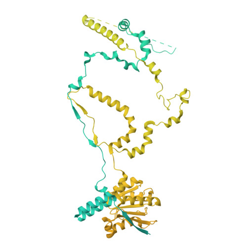
Helicase SRCAP3,230Homo sapiensMutation(s): 0 
EC: 3.6.4
UniProt & NIH Common Fund Data Resources
Find proteins for Q6ZRS2 (Homo sapiens)
Explore Q6ZRS2 
Go to UniProtKB:  Q6ZRS2
PHAROS:  Q6ZRS2
GTEx:  ENSG00000080603 
Entity Groups
Sequence Clusters30% Identity50% Identity70% Identity90% Identity95% Identity100% Identity
UniProt GroupQ6ZRS2
Sequence Annotations
Expand
Reference Sequence
Find similar proteins by:|  3D Structure
Entity ID: 2
MoleculeChains  Sequence LengthOrganismDetailsImage
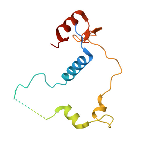
Vacuolar protein sorting-associated protein 72 homolog364Homo sapiensMutation(s): 0 
UniProt & NIH Common Fund Data Resources
Find proteins for Q15906 (Homo sapiens)
Explore Q15906 
Go to UniProtKB:  Q15906
PHAROS:  Q15906
GTEx:  ENSG00000163159 
Entity Groups
Sequence Clusters30% Identity50% Identity70% Identity90% Identity95% Identity100% Identity
UniProt GroupQ15906
Sequence Annotations
Expand
Reference Sequence
Find similar proteins by:|  3D Structure
Entity ID: 3
MoleculeChains  Sequence LengthOrganismDetailsImage
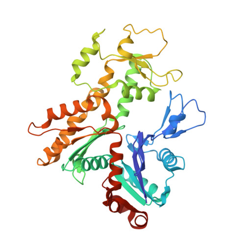
Actin-related protein 6396Homo sapiensMutation(s): 0 
UniProt & NIH Common Fund Data Resources
Find proteins for Q9GZN1 (Homo sapiens)
Explore Q9GZN1 
Go to UniProtKB:  Q9GZN1
PHAROS:  Q9GZN1
GTEx:  ENSG00000075089 
Entity Groups
Sequence Clusters30% Identity50% Identity70% Identity90% Identity95% Identity100% Identity
UniProt GroupQ9GZN1
Sequence Annotations
Expand
Reference Sequence
Find similar proteins by:|  3D Structure
Entity ID: 4
MoleculeChains  Sequence LengthOrganismDetailsImage
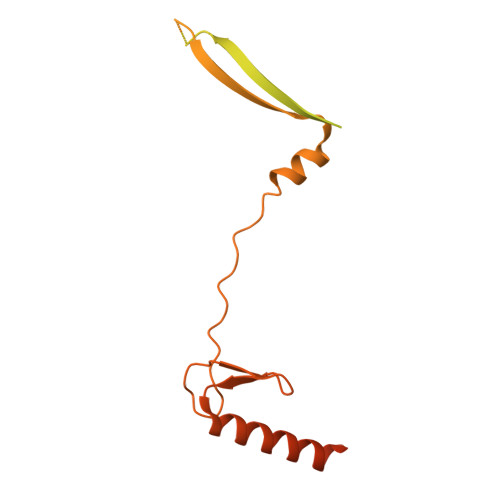
Zinc finger HIT domain-containing protein 1154Homo sapiensMutation(s): 0 
UniProt & NIH Common Fund Data Resources
Find proteins for O43257 (Homo sapiens)
Explore O43257 
Go to UniProtKB:  O43257
PHAROS:  O43257
GTEx:  ENSG00000106400 
Entity Groups
Sequence Clusters30% Identity50% Identity70% Identity90% Identity95% Identity100% Identity
UniProt GroupO43257
Sequence Annotations
Expand
Reference Sequence
Find similar proteins by:|  3D Structure
Entity ID: 5
MoleculeChains  Sequence LengthOrganismDetailsImage
RuvB-like 1
E, G, I
456Homo sapiensMutation(s): 0 
EC: 3.6.4.12
UniProt & NIH Common Fund Data Resources
Find proteins for Q9Y265 (Homo sapiens)
Explore Q9Y265 
Go to UniProtKB:  Q9Y265
PHAROS:  Q9Y265
GTEx:  ENSG00000175792 
Entity Groups
Sequence Clusters30% Identity50% Identity70% Identity90% Identity95% Identity100% Identity
UniProt GroupQ9Y265
Sequence Annotations
Expand
Reference Sequence
Find similar proteins by:|  3D Structure
Entity ID: 6
MoleculeChains  Sequence LengthOrganismDetailsImage
RuvB-like 2
F, H, J
463Homo sapiensMutation(s): 0 
EC: 3.6.4.12
UniProt & NIH Common Fund Data Resources
Find proteins for Q9Y230 (Homo sapiens)
Explore Q9Y230 
Go to UniProtKB:  Q9Y230
PHAROS:  Q9Y230
GTEx:  ENSG00000183207 
Entity Groups
Sequence Clusters30% Identity50% Identity70% Identity90% Identity95% Identity100% Identity
UniProt GroupQ9Y230
Sequence Annotations
Expand
Reference Sequence

Small Molecules

Ligands 4 Unique
IDChains Name / Formula / InChI Key2D Diagram3D Interactions
AGS

Query on AGS


Download:Ideal Coordinates CCD File
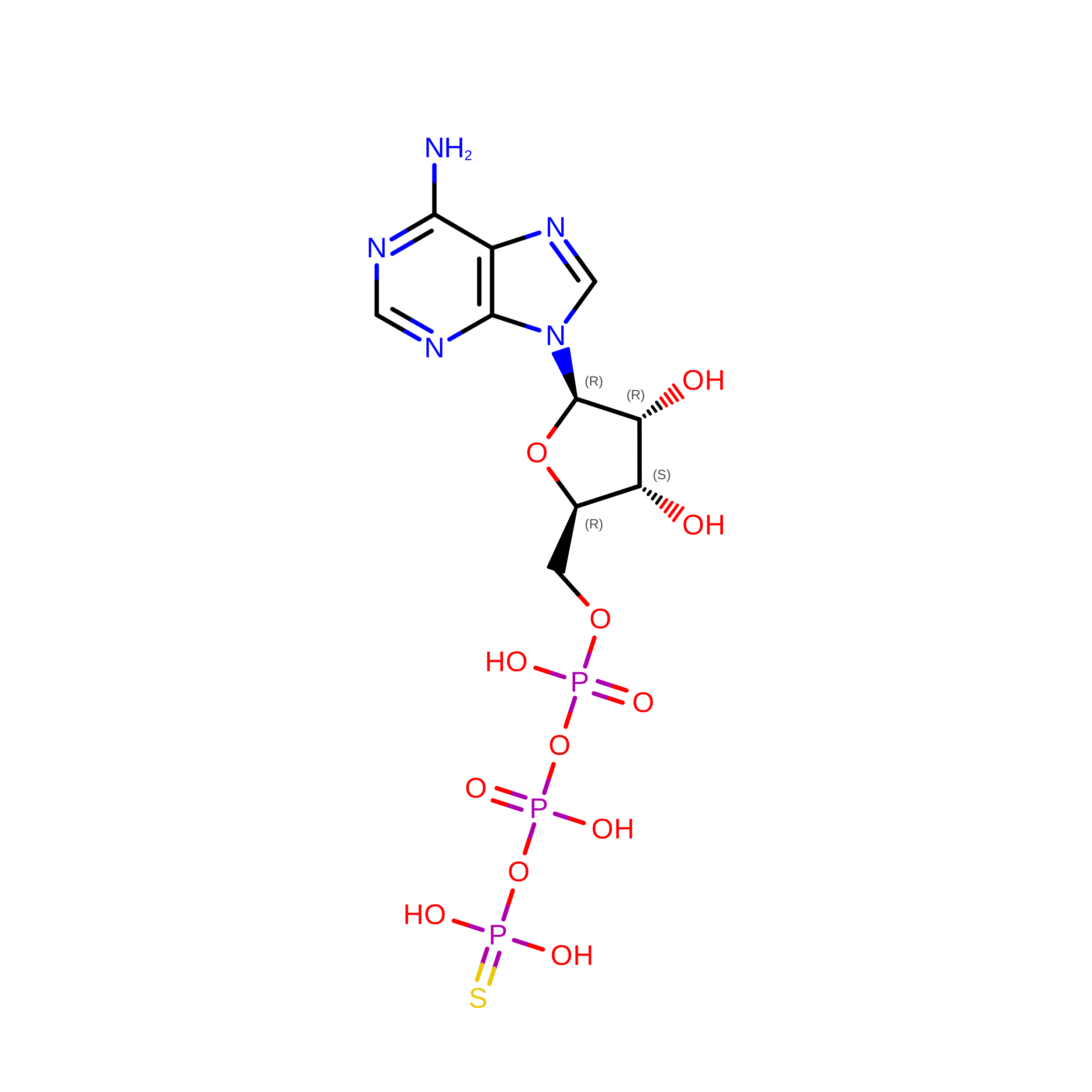
K [auth C]PHOSPHOTHIOPHOSPHORIC ACID-ADENYLATE ESTER
C10 H16 N5 O12 P3 S
NLTUCYMLOPLUHL-KQYNXXCUSA-N
ADP

Query on ADP


Download:Ideal Coordinates CCD File
O [auth E]
P [auth F]
R [auth G]
S [auth H]
U [auth I]
O [auth E],
P [auth F],
R [auth G],
S [auth H],
U [auth I],
V [auth J]
ADENOSINE-5'-DIPHOSPHATE
C10 H15 N5 O10 P2
XTWYTFMLZFPYCI-KQYNXXCUSA-N
ZN

Query on ZN


Download:Ideal Coordinates CCD File
M [auth D],
N [auth D]
ZINC ION
Zn
PTFCDOFLOPIGGS-UHFFFAOYSA-N
MG

Query on MG


Download:Ideal Coordinates CCD File
L [auth C],
Q [auth F],
T [auth H],
W [auth J]
MAGNESIUM ION
Mg
JLVVSXFLKOJNIY-UHFFFAOYSA-N

Experimental Data & Validation

Experimental Data

  • Method: ELECTRON MICROSCOPY
  • Resolution: 3.56 Å
  • Aggregation State: PARTICLE 
  • Reconstruction Method: SINGLE PARTICLE 
EM Software:
TaskSoftware PackageVersion
MODEL REFINEMENTPHENIX1.21_5207

Structure Validation

View Full Validation Report


Entry History 

& Funding Information

Deposition Data


Funding OrganizationLocationGrant Number
National Institutes of Health/National Institute of General Medical Sciences (NIH/NIGMS)United States--

Revision History  (Full details and data files)

  • Version 1.0: 2025-06-18
    Type: Initial release
  • Version 1.1: 2026-08-12
    Changes: Data collection, Database references