9BS3

Wild type DNA Ligase 1 with 5'-rG:C


Experimental Data Snapshot

  • Method: X-RAY DIFFRACTION
  • Resolution: 2.69 Å
  • R-Value Free: 0.249 
  • R-Value Work: 0.208 
  • R-Value Observed: 0.210 

Starting Model: experimental
View more details

wwPDB Validation   3D Report Full Report


Ligand Structure Quality Assessment 


This is version 1.0 of the entry. See complete history


Literature

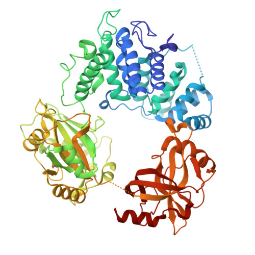
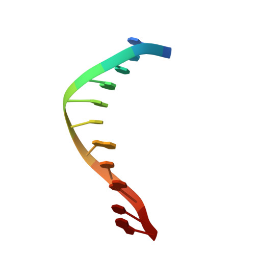
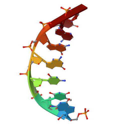
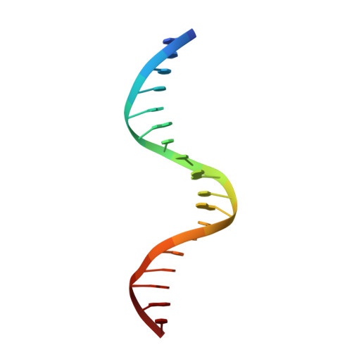
Structures of LIG1 uncover the mechanism of sugar discrimination against 5'-RNA-DNA junctions during ribonucleotide excision repair.

Balu, K.E.Tang, Q.Almohdar, D.Ratcliffe, J.Kalaycioglu, M.Caglayan, M.

(2024) J Biol Chem 300: 107688-107688

  • DOI: https://doi.org/10.1016/j.jbc.2024.107688
  • Primary Citation of Related Structures:  
    9BS3, 9BS4

  • PubMed Abstract: 

    Ribonucleotides in DNA cause several types of genome instability and can be removed by ribonucleotide excision repair (RER) that is finalized by DNA ligase 1 (LIG1). However, the mechanism by which LIG1 discriminates the RER intermediate containing a 5'-RNA-DNA lesion generated by RNase H2-mediated cleavage of ribonucleotides at atomic resolution remains unknown. Here, we determine X-ray structures of LIG1/5'-rG:C at the initial step of ligation where AMP is bound to the active site of the ligase and uncover a large conformational change downstream the nick resulting in a shift at Arg(R)871 residue in the Adenylation domain of the ligase. Furthermore, we demonstrate a diminished ligation of the nick DNA substrate with a 5'-ribonucleotide in comparison to an efficient end joining of the nick substrate with a 3'-ribonucleotide by LIG1. Finally, our results demonstrate that mutations at the active site residues of the ligase and LIG1 disease-associated variants significantly impact the ligation efficiency of RNA-DNA heteroduplexes harboring "wrong" sugar at 3'- or 5'-end of nick. Collectively, our findings provide a novel atomic insight into proficient sugar discrimination by LIG1 during the processing of the most abundant form of DNA damage in cells, genomic ribonucleotides, during the initial step of the RER pathway.


  • Organizational Affiliation

    Department of Biochemistry and Molecular Biology, University of Florida, Gainesville, Florida, USA.


Macromolecules

Find similar proteins by:  (by identity cutoff)  |  3D Structure
Entity ID: 1
MoleculeChains Sequence LengthOrganismDetailsImage
DNA ligase 1
A, E
644Homo sapiensMutation(s): 0 
Gene Names: LIG1
EC: 6.5.1.1
UniProt & NIH Common Fund Data Resources
Find proteins for P18858 (Homo sapiens)
Explore P18858 
Go to UniProtKB:  P18858
PHAROS:  P18858
GTEx:  ENSG00000105486 
Entity Groups  
Sequence Clusters30% Identity50% Identity70% Identity90% Identity95% Identity100% Identity
UniProt GroupP18858
Sequence Annotations
Expand
  • Reference Sequence

Find similar nucleic acids by:  Sequence   |   3D Structure  

Entity ID: 2
MoleculeChains LengthOrganismImage
DNA (5'-D(*GP*CP*TP*GP*AP*TP*GP*CP*GP*TP*G)-3')
B, F
11synthetic construct
Sequence Annotations
Expand
  • Reference Sequence

Find similar nucleic acids by:  Sequence   |   3D Structure  

Entity ID: 3
MoleculeChains LengthOrganismImage
DNA/RNA (5'-R(P*G)-D(P*TP*CP*GP*GP*AP*C)-3')
C, G
7synthetic construct
Sequence Annotations
Expand
  • Reference Sequence

Find similar nucleic acids by:  Sequence   |   3D Structure  

Entity ID: 4
MoleculeChains LengthOrganismImage
DNA (5'-D(P*GP*TP*CP*CP*GP*AP*CP*CP*AP*CP*GP*CP*AP*TP*CP*AP*GP*C)-3')
D, H
18synthetic construct
Sequence Annotations
Expand
  • Reference Sequence
Small Molecules
Ligands 1 Unique
IDChains Name / Formula / InChI Key2D Diagram3D Interactions
AMP (Subject of Investigation/LOI)
Query on AMP

Download Ideal Coordinates CCD File 
I [auth A],
J [auth E]
ADENOSINE MONOPHOSPHATE
C10 H14 N5 O7 P
UDMBCSSLTHHNCD-KQYNXXCUSA-N
Experimental Data & Validation

Experimental Data

  • Method: X-RAY DIFFRACTION
  • Resolution: 2.69 Å
  • R-Value Free: 0.249 
  • R-Value Work: 0.208 
  • R-Value Observed: 0.210 
  • Space Group: P 1 21 1
Unit Cell:
Length ( Å )Angle ( ˚ )
a = 71.348α = 90
b = 117.287β = 98.82
c = 104.048γ = 90
Software Package:
Software NamePurpose
PHENIXrefinement
HKL-2000data reduction
HKL-2000data scaling
PHASERphasing

Structure Validation

View Full Validation Report


Ligand Structure Quality Assessment 


Entry History & Funding Information

Deposition Data


Funding OrganizationLocationGrant Number
National Institutes of Health/National Institute of General Medical Sciences (NIH/NIGMS)United States1R35GM14711101

Revision History  (Full details and data files)

  • Version 1.0: 2024-09-25
    Type: Initial release