9B9O

Crystal structure of FlcD from Pseudomonas aeruginosa bond to iron(II) and substrate


Experimental Data Snapshot

  • Method: X-RAY DIFFRACTION
  • Resolution: 2.16 Å
  • R-Value Free: 0.233 
  • R-Value Work: 0.194 
  • R-Value Observed: 0.196 

Starting Model: experimental
View more details

wwPDB Validation   3D Report Full Report


Ligand Structure Quality Assessment 


This is version 1.2 of the entry. See complete history


Literature

Structural Basis for Methine Excision by a Heme Oxygenase-like Enzyme.

Simke, W.C.Walker, M.E.Calderone, L.A.Putz, A.T.Patteson, J.B.Vitro, C.N.Zizola, C.F.Redinbo, M.R.Pandelia, M.E.Grove, T.L.Li, B.

(2024) ACS Cent Sci 10: 1524-1536

  • DOI: https://doi.org/10.1021/acscentsci.4c00015
  • Primary Citation of Related Structures:  
    8W1Q, 9B9M, 9B9N, 9B9O

  • PubMed Abstract: 

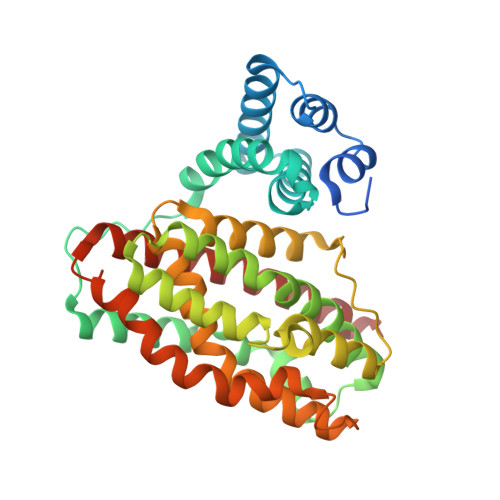
    Heme oxygenase-like domain-containing oxidases (HDOs) are a rapidly expanding enzyme family that typically use dinuclear metal cofactors instead of heme. FlcD, an HDO from the opportunistic pathogen Pseudomonas aeruginosa , catalyzes the excision of an oxime carbon in the biosynthesis of the copper-containing antibiotic fluopsin C. We show that FlcD is a dioxygenase that catalyzes a four-electron oxidation. Crystal structures of FlcD reveal a mononuclear iron in the active site, which is coordinated by two histidines, one glutamate, and the oxime of the substrate. Enzyme activity, Mössbauer spectroscopy, and electron paramagnetic resonance spectroscopy analyses support the usage of a mononuclear iron cofactor. This cofactor resembles that of mononuclear non-heme iron-dependent enzymes and breaks the paradigm of dinuclear HDO cofactors. This study begins to illuminate the catalytic mechanism of methine excision and indicates convergent evolution of different lineages of mononuclear iron-dependent enzymes.


  • Organizational Affiliation

    Department of Chemistry, The University of North Carolina at Chapel Hill, Chapel Hill, North Carolina 27599, United States.


Macromolecules
Find similar proteins by:  (by identity cutoff)  |  3D Structure
Entity ID: 1
MoleculeChains Sequence LengthOrganismDetailsImage
Pyrroloquinoline quinone (Coenzyme PQQ) biosynthesis protein C
A, B
351Pseudomonas aeruginosaMutation(s): 0 
Gene Names: pqqC_2CAZ10_19980DT376_18990NCTC13621_03444PAERUG_P19_London_7_VIM_2_05_10_05324
EC: 1.3.3.11
UniProt
Find proteins for A0A0C7AN42 (Pseudomonas aeruginosa)
Explore A0A0C7AN42 
Go to UniProtKB:  A0A0C7AN42
Entity Groups  
Sequence Clusters30% Identity50% Identity70% Identity90% Identity95% Identity100% Identity
UniProt GroupA0A0C7AN42
Sequence Annotations
Expand
  • Reference Sequence
Experimental Data & Validation

Experimental Data

  • Method: X-RAY DIFFRACTION
  • Resolution: 2.16 Å
  • R-Value Free: 0.233 
  • R-Value Work: 0.194 
  • R-Value Observed: 0.196 
  • Space Group: C 1 2 1
Unit Cell:
Length ( Å )Angle ( ˚ )
a = 124.991α = 90
b = 55.849β = 96.8
c = 96.558γ = 90
Software Package:
Software NamePurpose
PHENIXrefinement
XDSdata reduction
XDSdata scaling
PHASERphasing

Structure Validation

View Full Validation Report


Ligand Structure Quality Assessment 


Entry History & Funding Information

Deposition Data


Funding OrganizationLocationGrant Number
National Institutes of Health/National Institute of General Medical Sciences (NIH/NIGMS)United StatesGM148685
National Institutes of Health/National Institute of General Medical Sciences (NIH/NIGMS)United StatesGM137286

Revision History  (Full details and data files)

  • Version 1.0: 2024-07-31
    Type: Initial release
  • Version 1.1: 2024-09-11
    Changes: Database references
  • Version 1.2: 2024-09-18
    Changes: Database references