9B8U

Crystal structure of CRX-Ret4 oligonucleotide complex


Experimental Data Snapshot

  • Method: X-RAY DIFFRACTION
  • Resolution: 2.90 Å
  • R-Value Free: 0.249 
  • R-Value Work: 0.249 
  • R-Value Observed: 0.253 

Starting Model: experimental
View more details

wwPDB Validation   3D Report Full Report


This is version 1.1 of the entry. See complete history


Literature

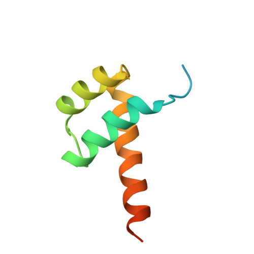
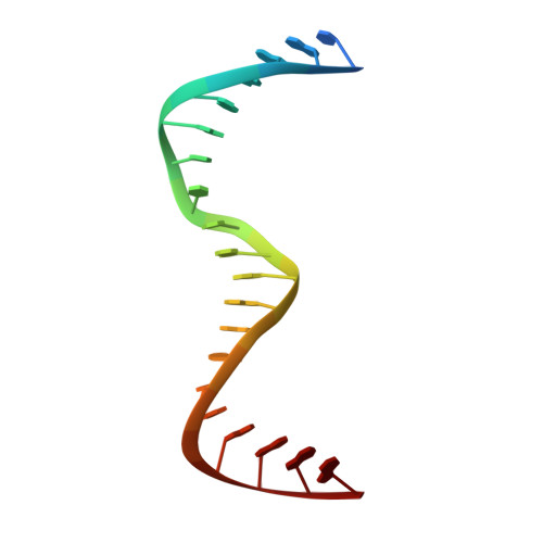
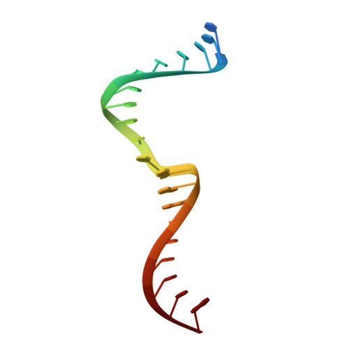
Molecular basis of CRX/DNA recognition and stoichiometry at the Ret4 response element.

Srivastava, D.Gowribidanur-Chinnaswamy, P.Gaur, P.Spies, M.Swaroop, A.Artemyev, N.O.

(2024) Structure 

  • DOI: https://doi.org/10.1016/j.str.2024.07.004
  • Primary Citation of Related Structures:  
    9B8U

  • PubMed Abstract: 

    Two retinal transcription factors, cone-rod homeobox (CRX) and neural retina leucine zipper (NRL), cooperate functionally and physically to control photoreceptor development and homeostasis. Mutations in CRX and NRL cause severe retinal diseases. Despite the roles of NRL and CRX, insight into their functions at the molecular level is lacking. Here, we have solved the crystal structure of the CRX homeodomain in complex with its cognate response element (Ret4) from the rhodopsin proximal promoter region. The structure reveals an unexpected 2:1 stoichiometry of CRX/Ret4 and unique orientation of CRX molecules on DNA, and it explains the mechanisms of pathogenic mutations in CRX. Mutations R41Q and E42K disrupt the CRX protein-protein contacts based on the structure and reduce the CRX/Ret4 binding stoichiometry, suggesting a novel disease mechanism. Furthermore, we show that NRL alters the stoichiometry and increases affinity of CRX binding at the rhodopsin promoter, which may enhance transcription of rod-specific genes and suppress transcription of cone-specific genes.


  • Organizational Affiliation

    Department of Molecular Physiology and Biophysics, The University of Iowa Carver College of Medicine, Iowa City, IA 52242, USA.


Macromolecules

Find similar proteins by:  (by identity cutoff)  |  3D Structure
Entity ID: 1
MoleculeChains Sequence LengthOrganismDetailsImage
Cone-rod homeobox protein
A, B, C, D
81Homo sapiensMutation(s): 0 
Gene Names: CRXCORD2
UniProt & NIH Common Fund Data Resources
Find proteins for O43186 (Homo sapiens)
Explore O43186 
Go to UniProtKB:  O43186
PHAROS:  O43186
GTEx:  ENSG00000105392 
Entity Groups  
Sequence Clusters30% Identity50% Identity70% Identity90% Identity95% Identity100% Identity
UniProt GroupO43186
Sequence Annotations
Expand
  • Reference Sequence

Find similar nucleic acids by:  Sequence   |   3D Structure  

Entity ID: 2
MoleculeChains LengthOrganismImage
DNA (5'-D(P*CP*AP*GP*GP*AP*GP*CP*TP*TP*AP*GP*GP*AP*GP*GP*GP*GP*GP*AP*G)-3')
E, G
21Homo sapiens
Sequence Annotations
Expand
  • Reference Sequence

Find similar nucleic acids by:  Sequence   |   3D Structure  

Entity ID: 3
MoleculeChains LengthOrganismImage
DNA (5'-D(P*CP*TP*CP*CP*CP*CP*CP*TP*CP*CP*TP*AP*AP*GP*CP*TP*CP*CP*TP*G)-3')
F, H
21Homo sapiens
Sequence Annotations
Expand
  • Reference Sequence
Small Molecules
Ligands 5 Unique
IDChains Name / Formula / InChI Key2D Diagram3D Interactions
CAC
Query on CAC

Download Ideal Coordinates CCD File 
I [auth A],
L [auth E]
CACODYLATE ION
C2 H6 As O2
OGGXGZAMXPVRFZ-UHFFFAOYSA-M
MPD
Query on MPD

Download Ideal Coordinates CCD File 
J [auth A](4S)-2-METHYL-2,4-PENTANEDIOL
C6 H14 O2
SVTBMSDMJJWYQN-YFKPBYRVSA-N
GOL
Query on GOL

Download Ideal Coordinates CCD File 
K [auth B],
N [auth F],
R [auth H],
S [auth H]
GLYCEROL
C3 H8 O3
PEDCQBHIVMGVHV-UHFFFAOYSA-N
K
Query on K

Download Ideal Coordinates CCD File 
M [auth E],
O [auth F],
T [auth H]
POTASSIUM ION
K
NPYPAHLBTDXSSS-UHFFFAOYSA-N
NA
Query on NA

Download Ideal Coordinates CCD File 
P [auth G],
Q [auth G]
SODIUM ION
Na
FKNQFGJONOIPTF-UHFFFAOYSA-N
Experimental Data & Validation

Experimental Data

  • Method: X-RAY DIFFRACTION
  • Resolution: 2.90 Å
  • R-Value Free: 0.249 
  • R-Value Work: 0.249 
  • R-Value Observed: 0.253 
  • Space Group: C 1 2 1
Unit Cell:
Length ( Å )Angle ( ˚ )
a = 109.597α = 90
b = 75.494β = 120.248
c = 97.229γ = 90
Software Package:
Software NamePurpose
PHENIXrefinement
XDSdata reduction
XSCALEdata scaling
PHASERphasing

Structure Validation

View Full Validation Report


Entry History & Funding Information

Deposition Data


Funding OrganizationLocationGrant Number
National Institutes of Health/National Eye Institute (NIH/NEI)United States1R01EY034455-01

Revision History  (Full details and data files)

  • Version 1.0: 2024-07-31
    Type: Initial release
  • Version 1.1: 2024-08-14
    Changes: Database references