9B2H

HIV-1 wild type protease with GRL-072-17A, a substituted tetrahydrofuran derivative based on Darunavir as P2 group

  • Classification: VIRAL PROTEIN
  • Organism(s): Human immunodeficiency virus 1
  • Expression System: Escherichia coli BL21(DE3)
  • Mutation(s): Yes 

  • Deposited: 2024-03-15 Released: 2024-07-24 
  • Deposition Author(s): Wang, Y.-F., Agniswamy, J., Ghosh, A.K., Weber, I.T.
  • Funding Organization(s): National Institutes of Health/National Institute Of Allergy and Infectious Diseases (NIH/NIAID), Department of Energy (DOE, United States), Japan Agency for Medical Research and Development (AMED), National Institutes of Health/National Cancer Institute (NIH/NCI), National Center for Global Health and Medicine (NCGM) Research Institute, Welfare and Labor of Japan, Grant-in-Aid for Scientific Research and a Grant-in-Aid for Challenging Research from the Ministry of Education, Culture, Sports, Science, and Technology of Japan (Monbu Kagakusho)

Experimental Data Snapshot

  • Method: X-RAY DIFFRACTION
  • Resolution: 1.32 Å
  • R-Value Free: 0.164 
  • R-Value Work: 0.130 
  • R-Value Observed: 0.132 

Starting Model: experimental
View more details

wwPDB Validation   3D Report Full Report


Ligand Structure Quality Assessment 


This is version 1.0 of the entry. See complete history


Literature

Design of substituted tetrahydrofuran derivatives for HIV-1 protease inhibitors: synthesis, biological evaluation, and X-ray structural studies.

Ghosh, A.K.Lee, D.Sharma, A.Johnson, M.E.Ghosh, A.K.Wang, Y.F.Agniswamy, J.Amano, M.Hattori, S.I.Weber, I.T.Mitsuya, H.

(2024) Org Biomol Chem 

  • DOI: https://doi.org/10.1039/d4ob00506f
  • Primary Citation of Related Structures:  
    9B2H

  • PubMed Abstract: 

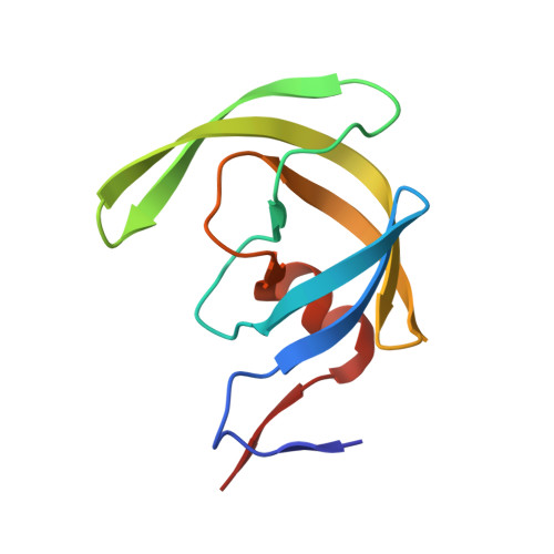
    Substituted tetrahydrofuran derivatives were designed and synthesized to serve as the P2 ligand for a series of potent HIV-1 protease inhibitors. Both enantiomers of the tetrahydrofuran derivatives were synthesized stereoselectivity in optically active forms using lipase-PS catalyzed enzymatic resolution as the key step. These tetrahydrofuran derivatives are designed to promote hydrogen bonding and van der Waals interactions with the backbone atoms in the S2 subsite of the HIV-1 protease active site. Several inhibitors displayed very potent HIV-1 protease inhibitory activity. A high-resolution X-ray crystal structure of an inhibitor-bound HIV-1 protease provided important insight into the ligand binding site interactions in the active site.


  • Organizational Affiliation

    Department of Chemistry, Purdue University, 560 Oval Drive, West Lafayette, IN 47907, USA. akghosh@purdue.edu.


Macromolecules
Find similar proteins by:  (by identity cutoff)  |  3D Structure
Entity ID: 1
MoleculeChains Sequence LengthOrganismDetailsImage
Protease
A, B
99Human immunodeficiency virus 1Mutation(s): 5 
Gene Names: pol
EC: 3.4.23.16
UniProt
Find proteins for Q5RZ08 (Human immunodeficiency virus 1)
Explore Q5RZ08 
Go to UniProtKB:  Q5RZ08
Entity Groups  
Sequence Clusters30% Identity50% Identity70% Identity90% Identity95% Identity100% Identity
UniProt GroupQ5RZ08
Sequence Annotations
Expand
  • Reference Sequence
Small Molecules
Ligands 5 Unique
IDChains Name / Formula / InChI Key2D Diagram3D Interactions
A1AIH (Subject of Investigation/LOI)
Query on A1AIH

Download Ideal Coordinates CCD File 
H [auth B]2,5:6,9-dianhydro-1,3,7,8-tetradeoxy-4-O-({(2S,3R)-3-hydroxy-4-[(4-methoxybenzene-1-sulfonyl)(2-methylpropyl)amino]-1-phenylbutan-2-yl}carbamoyl)-L-gluco-nonitol
C31 H44 N2 O8 S
HNDYYNUFXXLWEK-LDAKCSIQSA-N
GOL
Query on GOL

Download Ideal Coordinates CCD File 
F [auth A],
L [auth B]
GLYCEROL
C3 H8 O3
PEDCQBHIVMGVHV-UHFFFAOYSA-N
FMT
Query on FMT

Download Ideal Coordinates CCD File 
G [auth A]FORMIC ACID
C H2 O2
BDAGIHXWWSANSR-UHFFFAOYSA-N
CL
Query on CL

Download Ideal Coordinates CCD File 
D [auth A],
E [auth A],
J [auth B],
K [auth B]
CHLORIDE ION
Cl
VEXZGXHMUGYJMC-UHFFFAOYSA-M
NA
Query on NA

Download Ideal Coordinates CCD File 
C [auth A],
I [auth B]
SODIUM ION
Na
FKNQFGJONOIPTF-UHFFFAOYSA-N
Experimental Data & Validation

Experimental Data

  • Method: X-RAY DIFFRACTION
  • Resolution: 1.32 Å
  • R-Value Free: 0.164 
  • R-Value Work: 0.130 
  • R-Value Observed: 0.132 
  • Space Group: P 21 21 2
Unit Cell:
Length ( Å )Angle ( ˚ )
a = 58.576α = 90
b = 85.877β = 90
c = 46.312γ = 90
Software Package:
Software NamePurpose
REFMACrefinement
HKL-2000data scaling
HKL-2000data reduction
PHASERphasing
PDB_EXTRACTdata extraction

Structure Validation

View Full Validation Report


Ligand Structure Quality Assessment 


Entry History & Funding Information

Deposition Data


Funding OrganizationLocationGrant Number
National Institutes of Health/National Institute Of Allergy and Infectious Diseases (NIH/NIAID)United StatesAI150466
National Institutes of Health/National Institute Of Allergy and Infectious Diseases (NIH/NIAID)United StatesAI150461
Department of Energy (DOE, United States)United StatesW-31-109-Eng-38
Japan Agency for Medical Research and Development (AMED)JapanJP15fk0410001, JP16fk0410101
National Institutes of Health/National Cancer Institute (NIH/NCI)United States--
National Center for Global Health and Medicine (NCGM) Research InstituteJapan--
Welfare and Labor of JapanJapan--
Grant-in-Aid for Scientific Research and a Grant-in-Aid for Challenging Research from the Ministry of Education, Culture, Sports, Science, and Technology of Japan (Monbu Kagakusho)Japan--

Revision History  (Full details and data files)

  • Version 1.0: 2024-07-24
    Type: Initial release