Structure-driven development of a biomimetic rare earth artificial metalloprotein.
Thompson, P.J., Boggs, D.G., Wilson, C.A., Bruchs, A.T., Velidandla, U., Bridwell-Rabb, J., Olshansky, L.(2024) Proc Natl Acad Sci U S A 121: e2405836121-e2405836121
- PubMed: 39116128 Search on PubMedSearch on PubMed Central
- DOI: https://doi.org/10.1073/pnas.2405836121
- Primary Citation Related Structures:
9B1U, 9B1V - PubMed Abstract:
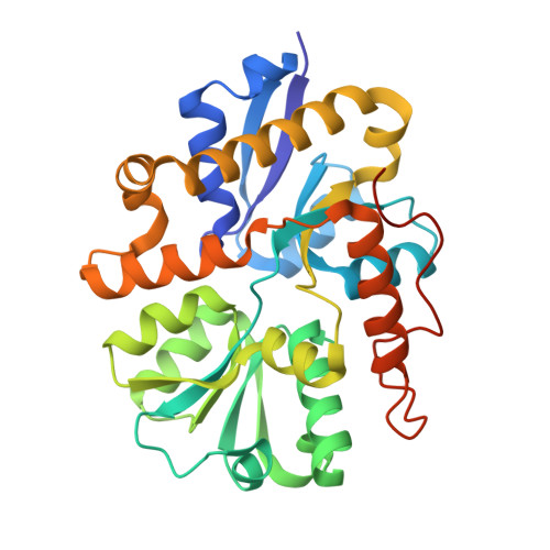
The 2011 discovery of the first rare earth-dependent enzyme in methylotrophic Methylobacterium extorquens AM1 prompted intensive research toward understanding the unique chemistry at play in these systems. This enzyme, an alcohol dehydrogenase (ADH), features a La 3+ ion closely associated with redox-active coenzyme pyrroloquinoline quinone (PQQ) and is structurally homologous to the Ca 2+ -dependent ADH from the same organism. AM1 also produces a periplasmic PQQ-binding protein, PqqT, which we have now structurally characterized to 1.46-Å resolution by X-ray diffraction. This crystal structure reveals a Lys residue hydrogen-bonded to PQQ at the site analogously occupied by a Lewis acidic cation in ADH. Accordingly, we prepared K 142 A- and K 142 D-PqqT variants to assess the relevance of this site toward metal binding. Isothermal titration calorimetry experiments and titrations monitored by UV-Vis absorption and emission spectroscopies support that K 142 D-PqqT binds tightly ( K d = 0.6 ± 0.2 μM) to La 3+ in the presence of bound PQQ and produces spectral signatures consistent with those of ADH enzymes. These spectral signatures are not observed for WT- or K 142 A-variants or upon addition of Ca 2+ to PQQ ⸦ K 142 D-PqqT. Addition of benzyl alcohol to La 3+ -bound PQQ ⸦ K 142 D-PqqT (but not Ca 2+ -bound PQQ ⸦ K 142 D-PqqT, or La 3+ -bound PQQ ⸦ WT-PqqT) produces spectroscopic changes associated with PQQ reduction, and chemical trapping experiments reveal the production of benzaldehyde, supporting ADH activity. By creating a metal binding site that mimics native ADH enzymes, we present a rare earth-dependent artificial metalloenzyme primed for future mechanistic, biocatalytic, and biosensing applications.
- Center for Biophysics and Quantitative Biology, University of Illinois, Urbana-Champaign, Urbana, IL 61801.
Organizational Affiliation: