9AU0 | pdb_00009au0

Cryo-EM structure of the BW245C-bound prostaglandin D2 receptor (DP1)-Gs complex


Experimental Data Snapshot

  • Method: ELECTRON MICROSCOPY
  • Resolution: 2.45 Å
  • Aggregation State: PARTICLE 
  • Reconstruction Method: SINGLE PARTICLE 

wwPDB Validation 3D Report Full Report

Validation slider image for 9AU0

This is version 1.1 of the entry. See complete history

Literature

Structural insights into the mechanism of activation and inhibition of the prostaglandin D2 receptor 1.

Davoudinasab, B.Raskovalov, A.Lee, W.Kim, D.Kim, H.Lam, J.H.Han, G.W.Katritch, V.Cherezov, V.

(2025) Nat Commun 16: 8944-8944

  • DOI: https://doi.org/10.1038/s41467-025-64002-z
  • Primary Citation Related Structures: 
    9AU0, 9E9S, 9EE5, 9EI5, 9EKH

  • PubMed Abstract: 

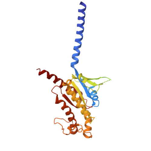
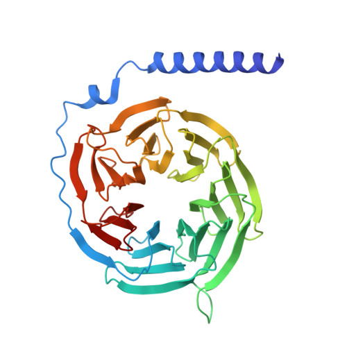
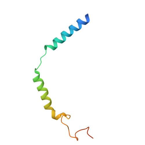
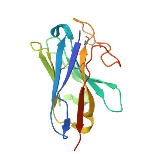
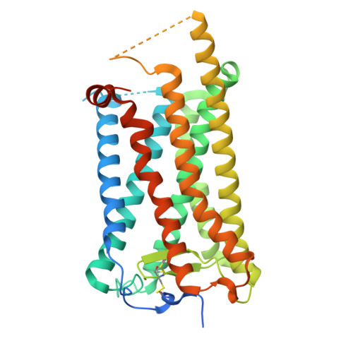
    The prostaglandin D2 receptor 1 (DP1), a member of the prostanoid G protein-coupled receptor (GPCR) family, plays critical roles in allergic responses, sleep regulation, immune modulation, and vasodilation. Here, we present five high-resolution cryo-electron microscopy (cryo-EM) structures of the human DP1 receptor, including an apo structure, two inactive state structures bound to two different inverse agonists developed by ONO Pharmaceutical, and two active state structures in complex with the G s protein and bound to the endogenous agonist PGD2 and its selective derivative BW245C. Structural analysis, complemented by molecular dynamics simulations and site-directed mutagenesis, reveals key residues involved in ligand recognition and suggests a distinct activation mechanism for DP1, which lacks most of the conserved class A GPCR motifs. Notably, the unique residue K76 within the conserved sodium pocket acts as a major activation switch, while amphiphilic helix 8 adopts an unconventional orientation essential for receptor function. These findings offer valuable insights into the structure and function of prostanoid receptors and may facilitate the development of therapeutics targeting DP1.


  • Organizational Affiliation
    • Department of Chemistry, University of Southern California, Los Angeles, CA, USA.

Macromolecule Content 

  • Total Structure Weight: 150.92 kDa 
  • Atom Count: 8,576 
  • Modeled Residue Count: 1,069 
  • Deposited Residue Count: 1,334 
  • Unique protein chains: 5

Macromolecules

Find similar proteins by:|  3D Structure
Entity ID: 1
MoleculeChains  Sequence LengthOrganismDetailsImage
Guanine nucleotide-binding protein G(s) subunit alpha isoforms short394Homo sapiensMutation(s): 8 
Gene Names: GNASGNAS1GSP
EC: 3.6.5
UniProt & NIH Common Fund Data Resources
Find proteins for P63092 (Homo sapiens)
Explore P63092 
Go to UniProtKB:  P63092
PHAROS:  P63092
GTEx:  ENSG00000087460 
Entity Groups
Sequence Clusters30% Identity50% Identity70% Identity90% Identity95% Identity100% Identity
UniProt GroupP63092
Sequence Annotations
Expand
Reference Sequence
Find similar proteins by:|  3D Structure
Entity ID: 2
MoleculeChains  Sequence LengthOrganismDetailsImage
Guanine nucleotide-binding protein G(I)/G(S)/G(T) subunit beta-1345Homo sapiensMutation(s): 0 
Gene Names: GNB1
UniProt
Find proteins for A0A8D2K0M5 (Theropithecus gelada)
Explore A0A8D2K0M5 
Go to UniProtKB:  A0A8D2K0M5
Entity Groups
Sequence Clusters30% Identity50% Identity70% Identity90% Identity95% Identity100% Identity
UniProt GroupA0A8D2K0M5
Sequence Annotations
Expand
Reference Sequence
Find similar proteins by:|  3D Structure
Entity ID: 3
MoleculeChains  Sequence LengthOrganismDetailsImage
Guanine nucleotide-binding protein G(I)/G(S)/G(O) subunit gamma-271Homo sapiensMutation(s): 0 
Gene Names: GNG2
UniProt & NIH Common Fund Data Resources
Find proteins for P59768 (Homo sapiens)
Explore P59768 
Go to UniProtKB:  P59768
PHAROS:  P59768
GTEx:  ENSG00000186469 
Entity Groups
Sequence Clusters30% Identity50% Identity70% Identity90% Identity95% Identity100% Identity
UniProt GroupP59768
Sequence Annotations
Expand
Reference Sequence
Find similar proteins by:|  3D Structure
Entity ID: 4
MoleculeChains  Sequence LengthOrganismDetailsImage
Nanobody-35D [auth N]139Lama glamaMutation(s): 0 
Find similar proteins by:|  3D Structure
Entity ID: 5
MoleculeChains  Sequence LengthOrganismDetailsImage
Prostaglandin D2 receptorE [auth R]385Homo sapiensMutation(s): 0 
Gene Names: PTGDR
UniProt & NIH Common Fund Data Resources
Find proteins for Q13258 (Homo sapiens)
Explore Q13258 
Go to UniProtKB:  Q13258
PHAROS:  Q13258
GTEx:  ENSG00000168229 
Entity Groups
Sequence Clusters30% Identity50% Identity70% Identity90% Identity95% Identity100% Identity
UniProt GroupQ13258
Glycosylation
Glycosylation Sites: 1Go to GlyGen: Q13258-1
Sequence Annotations
Expand
Reference Sequence

Small Molecules

Ligands 3 Unique
IDChains Name / Formula / InChI Key2D Diagram3D Interactions
Y01

Query on Y01


Download:Ideal Coordinates CCD File
H [auth R]CHOLESTEROL HEMISUCCINATE
C31 H50 O4
WLNARFZDISHUGS-MIXBDBMTSA-N
A1AF8
(Subject of Investigation/LOI)

Query on A1AF8


Download:Ideal Coordinates CCD File
F [auth R]7-{(4S)-3-[(3R)-3-cyclohexyl-3-hydroxypropyl]-2,5-dioxoimidazolidin-4-yl}heptanoic acid
C19 H32 N2 O5
ZIDQIOZJEJFMOH-JKSUJKDBSA-N
NAG

Query on NAG


Download:Ideal Coordinates CCD File
G [auth R]2-acetamido-2-deoxy-beta-D-glucopyranose
C8 H15 N O6
OVRNDRQMDRJTHS-FMDGEEDCSA-N

Experimental Data & Validation

Experimental Data

  • Method: ELECTRON MICROSCOPY
  • Resolution: 2.45 Å
  • Aggregation State: PARTICLE 
  • Reconstruction Method: SINGLE PARTICLE 
EM Software:
TaskSoftware PackageVersion
MODEL REFINEMENTPHENIX1.21_5207:
RECONSTRUCTIONcryoSPARC

Structure Validation

View Full Validation Report


Entry History 

& Funding Information

Deposition Data


Funding OrganizationLocationGrant Number
National Institutes of Health/National Institute of General Medical Sciences (NIH/NIGMS)United StatesR35GM127086

Revision History  (Full details and data files)

  • Version 1.0: 2025-08-27
    Type: Initial release
  • Version 1.1: 2026-03-11
    Changes: Data collection, Database references