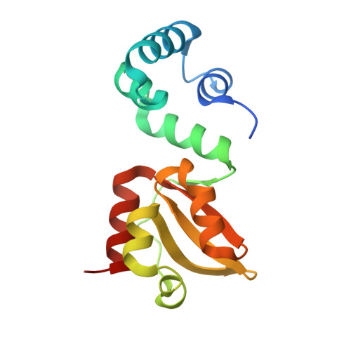
The structural basis for high-affinity c-di-GMP binding to the GSPII-B domain of the traffic ATPase PilF from Thermus thermophilus.
Neissner, K., Keller, H., Kirchner, L., Dusterhus, S., Duchardt-Ferner, E., Averhoff, B., Wohnert, J.(2024) J Biol Chem 301: 108041-108041
- PubMed: 39615687 Search on PubMed
- DOI: https://doi.org/10.1016/j.jbc.2024.108041
- Primary Citation Related Structures:
8PDK, 8PE0, 8PFA, 8PKZ, 8PQU - PubMed Abstract:
c-di-GMP is an important second messenger in bacteria regulating, for example motility, biofilm formation, cell wall biosynthesis, infectivity, and natural transformability. It binds to a multitude of intracellular receptors. This includes proteins containing general secretory pathway II (GSPII) domains such as the N-terminal domain of the Vibrio cholerae ATPase MshE (MshEN) which binds c-di-GMP with two copies of a 24-amino acids sequence motif. The traffic ATPase PilF from Thermus thermophilus is important for type IV pilus biogenesis, twitching motility, surface attachment, and natural DNA-uptake and contains three consecutive homologous GPSII domains. We show that only two of these domains bind c-di-GMP and define the structural basis for the exceptional high affinity of the GSPII-B domain for c-di-GMP, which is 83-fold higher than that of the prototypical MshEN domain. Our work establishes an extended consensus sequence for the c-di-GMP-binding motif and highlights the role of hydrophobic residues for high-affinity recognition of c-di-GMP. Our structure is the first example for a c-di-GMP-binding domain not relying on arginine residues for ligand recognition. We also show that c-di-GMP-binding induces local unwinding of an α-helical turn as well as subdomain reorientation to reinforce intermolecular contacts between c-di-GMP and the C-terminal subdomain. Abolishing c-di-GMP binding to GSPII-B reduces twitching motility and surface attachment but not natural DNA-uptake. Overall, our work contributes to a better characterization of c-di-GMP binding in this class of effector domains, allows the prediction of high-affinity c-di-GMP-binding family members, and advances our understanding of the importance of c-di-GMP binding for T4P-related functions.
- Institute for Molecular Biosciences, Goethe-University Frankfurt/M., Frankfurt, Germany; Center for Biomolecular Magnetic Resonance (BMRZ), Goethe-University Frankfurt/M., Frankfurt, Germany.
Organizational Affiliation: