8ZT4 | pdb_00008zt4

Complex structure of N-acetyltransferase SbzI and carrier protein SbzG in the biosynthesis of altemicidin


Experimental Data Snapshot

  • Method: X-RAY DIFFRACTION
  • Resolution: 2.60 Å
  • R-Value Free: 
    0.234 (Depositor), 0.244 (DCC) 
  • R-Value Work: 
    0.196 (Depositor) 
  • R-Value Observed: 
    0.198 (Depositor) 

Starting Model: in silico
View more details

wwPDB Validation 3D Report Full Report

Validation slider image for 8ZT4

This is version 1.0 of the entry. See complete history

Literature

Structure-function analysis of carrier protein-dependent 2-sulfamoylacetyl transferase in the biosynthesis of altemicidin.

Zhu, Y.Mori, T.Karasawa, M.Shirai, K.Cheng, W.Terada, T.Awakawa, T.Abe, I.

(2024) Nat Commun 15: 10896-10896

  • DOI: https://doi.org/10.1038/s41467-024-55265-z
  • Primary Citation Related Structures: 
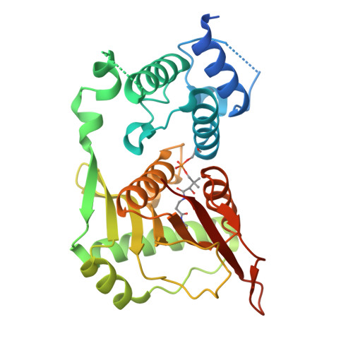
    8ZT3, 8ZT4

  • PubMed Abstract: 

    The general control non-repressible 5 (GCN5)-related N-acetyltransferase (GNAT) SbzI, in the biosynthesis of the sulfonamide antibiotic altemicidin, catalyzes the transfer of the 2-sulfamoylacetyl (2-SA) moiety onto 6-azatetrahydroindane dinucleotide. While most GNAT superfamily utilize acyl-coenzyme A (acyl-CoA) as substrates, SbzI recognizes a carrier-protein (CP)-tethered 2-SA substrate. Moreover, SbzI is the only naturally occurring enzyme that catalyzes the direct incorporation of sulfonamide, a valuable pharmacophore in medicinal chemistry. Here, we present the structure-function analysis and structure-based engineering of SbzI. The crystal structure of SbzI in complex with the CP SbzG, along with cross-linking and isothermal titration calorimetry analyses of their variants, revealed the structural basis for CP recognition by the GNAT SbzI. Furthermore, docking simulation, molecular dynamics simulation, and mutagenesis studies indicated the intimate structural details of the unique reaction mechanism of SbzI, which does not utilize a general base residue in contrast to other typical GNATs. These findings facilitated rational engineering of the enzyme to expand the substrate range and to generate azaindane dinucleotide derivatives.


  • Organizational Affiliation
    • Graduate School of Pharmaceutical Sciences, The University of Tokyo, Tokyo, Japan.

Macromolecule Content 

  • Total Structure Weight: 64.17 kDa 
  • Atom Count: 3,899 
  • Modeled Residue Count: 500 
  • Deposited Residue Count: 596 
  • Unique protein chains: 1

Macromolecules

Find similar proteins by:|  3D Structure
Entity ID: 1
MoleculeChains  Sequence LengthOrganismDetailsImage
Carrier protein,GNAT family transferase
A, B
298Streptomyces sp.Mutation(s): 0 
Gene Names: sbzGsbzI

Small Molecules

Ligands 3 Unique
IDChains Name / Formula / InChI Key2D Diagram3D Interactions
BCT

Query on BCT


Download:Ideal Coordinates CCD File
G [auth A],
H [auth A],
I [auth B],
J [auth B]
BICARBONATE ION
C H O3
BVKZGUZCCUSVTD-UHFFFAOYSA-M
SCN

Query on SCN


Download:Ideal Coordinates CCD File
C [auth A],
D [auth A],
E [auth A]
THIOCYANATE ION
C N S
ZMZDMBWJUHKJPS-UHFFFAOYSA-M
CL

Query on CL


Download:Ideal Coordinates CCD File
F [auth A],
K [auth B]
CHLORIDE ION
Cl
VEXZGXHMUGYJMC-UHFFFAOYSA-M
Modified Residues  1 Unique
IDChains TypeFormula2D DiagramParent
4HH
Query on 4HH
A, B
L-PEPTIDE LINKINGC14 H28 N3 O9 P SSER

Experimental Data & Validation

Experimental Data

  • Method: X-RAY DIFFRACTION
  • Resolution: 2.60 Å
  • R-Value Free:  0.234 (Depositor), 0.244 (DCC) 
  • R-Value Work:  0.196 (Depositor) 
  • R-Value Observed: 0.198 (Depositor) 
Space Group: P 21 21 21
Unit Cell:
Length ( Å )Angle ( ˚ )
a = 77.15α = 90
b = 95.68β = 90
c = 130.79γ = 90
Software Package:
Software NamePurpose
PHENIXrefinement
XDSdata reduction
XSCALEdata scaling
PHASERphasing

Structure Validation

View Full Validation Report


Entry History 

& Funding Information

Deposition Data


Funding OrganizationLocationGrant Number
Not funded--

Revision History  (Full details and data files)

  • Version 1.0: 2025-02-05
    Type: Initial release