Structural insight into sugar-binding modes of microbial ss-amylase.
Hirata, A., Mikami, B.(2024) Biochem Biophys Res Commun 733: 150695-150695
- PubMed: 39288698 Search on PubMed
- DOI: https://doi.org/10.1016/j.bbrc.2024.150695
- Primary Citation Related Structures:
8ZRZ - PubMed Abstract:
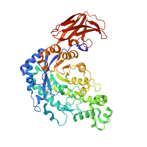
ß-Amylase, which catalyses the release of ß-anomeric maltose from the non-reducing end of starch, is widely used in the food industry. Increasing its enzyme activity through protein engineering might improve the efficiency of food processing. To obtain detailed structural information to assist rationale design, here the crystal structure of Bacillus cereus β-amylase (BCB) complexed with maltose was determined by molecular replacement and refined using anisotropic temperature factors to 1.26 Å resolution with R work /R free factors of 12.4/15.7 %. The structure contains six maltose and one glucose molecules, of which two maltose and one glucose are bound at sites not previously observed in BCB structures. These three new sugar-binding sites are located on the surface and likely to be important in enhancing the degradation of raw-starch granules. In the active site of BCB, two maltose molecules are bound in tandem at subsites -2 ∼ -1 and +1 ∼ +2. Notably, the conformation of the glucose moiety bound at subsite -1 is a mixture of α-anomeric distorted 1,4 B boat and 4 C 1 chair forms, while those at subsites -2, +1 ∼ +2 are all in the 4 C 1 chair forms. The O1 of the distorted α-glucose residue at subsite -1 occupies the position of the putative catalytic water, forming a hydrogen bond with OE1 of Glu367 (base catalyst), suggesting that this distorted sugar is not involved in catalysis. Together, these findings pave the way for further improving the functionality of microbial ß-amylase enzymes.
- Department of Natural Science, Graduate School of Technology, Industrial and Social Science, Tokushima University, 2-1 Minamijosanjimacho, Tokushima, Tokushima, 770-8506, Japan. Electronic address: ahirata@tokushima-u.ac.jp.
Organizational Affiliation: