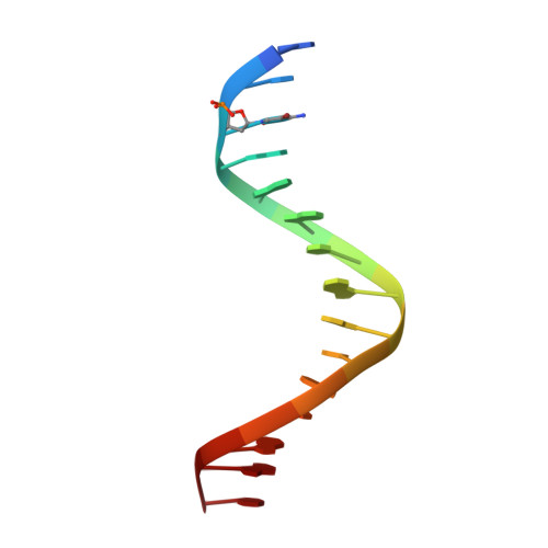
8WU5

The complex of CAG repeat sequence-specific binding cPIP and dsDNA with A-A mismatch


Experimental Data Snapshot

  • Method: X-RAY DIFFRACTION
  • Resolution: 2.80 Å
  • R-Value Free: 0.297 
  • R-Value Work: 0.284 
  • R-Value Observed: 0.285 

wwPDB Validation   3D Report Full Report


Ligand Structure Quality Assessment 


This is version 1.1 of the entry. See complete history


Literature

Structural Studies of a Complex of a CAG/CTG Repeat Sequence-Specific Binding Molecule and A-A-Mismatch-Containing DNA.

Abe, K.Hirose, Y.Kumagai, T.Hashiya, K.Hidaka, K.Emura, T.Bando, T.Takeda, K.Sugiyama, H.

(2024) JACS Au 4: 1801-1810

  • DOI: https://doi.org/10.1021/jacsau.3c00830
  • Primary Citation of Related Structures:  
    8WU5

  • PubMed Abstract: 

    Triplet repeat diseases are caused by the abnormal elongation of repeated sequences comprising three bases. In particular, the elongation of CAG/CTG repeat sequences is thought to result in conditions such as Huntington's disease and myotonic dystrophy type 1. Although the causes of these diseases are known, fundamental treatments have not been established, and specific drugs are expected to be developed. Pyrrole imidazole polyamide (PIP) is a class of molecules that binds to the minor groove of the DNA duplex in a sequence-specific manner; because of this property, it shows promise in drug discovery applications. Earlier, it was reported that PIP designed to bind CAG/CTG repeat sequences suppresses the genes that cause triplet repeat diseases. In this study, we performed an X-ray crystal structure analysis of a complex of double-stranded DNA containing A-A mismatched base pairs and a cyclic-PIP that binds specifically to CAG/CTG sequences. Furthermore, the validity and characteristics of this structure were analyzed using in silico molecular modeling, ab initio energy calculations, gel electrophoresis, and surface plasmon resonance. With our direct observation using atomic force microscopy and DNA origami, we revealed that the PIP caused structural changes in the DNA strands carrying the expanded CAG/CTG repeat. Overall, our study provides new insight into PIP from a structural perspective.


  • Organizational Affiliation

    Department of Chemistry, Graduate School of Science, Kyoto University, Sakyo, Kyoto 606-8502, Japan.


Macromolecules

Find similar nucleic acids by:  Sequence   |   3D Structure  

Entity ID: 1
MoleculeChains LengthOrganismImage
DNA (5'-D(*GP*CP*(CBR)P*GP*AP*GP*CP*AP*GP*CP*AP*CP*GP*GP*C)-3')
A, B, C, D
15synthetic construct
Sequence Annotations
Expand
  • Reference Sequence
Small Molecules
Ligands 1 Unique
IDChains Name / Formula / InChI Key2D Diagram3D Interactions
A1LYO
Query on A1LYO

Download Ideal Coordinates CCD File 
E [auth B],
F [auth C]
(1^2Z,4^2Z,11^2Z,14^2Z,22^2Z,25^2Z,32^2Z,35^2Z,19R,40R)-1^1,4^1,11^1,14^1,22^1,25^1,32^1,35^1-octamethyl-2,5,9,12,15,20,23,26,30,33,36,41-dodecaoxo-1^1H,4^1H,11^1H,14^1H,22^1H,25^1H,32^1H,35^1H-3,6,10,13,16,21,24,27,31,34,37,42-dodecaaza-1(2,4),11,22,32(4,2)-tetraimidazola-4,14,25,35(4,2)-tetrapyrrolacyclodotetracontaphane-19,40-diaminium
C58 H70 N26 O12
QKAIZNHPQCMMDA-KKLWWLSJSA-N
Experimental Data & Validation

Experimental Data

  • Method: X-RAY DIFFRACTION
  • Resolution: 2.80 Å
  • R-Value Free: 0.297 
  • R-Value Work: 0.284 
  • R-Value Observed: 0.285 
  • Space Group: P 21 21 21
Unit Cell:
Length ( Å )Angle ( ˚ )
a = 27.186α = 90
b = 44.051β = 90
c = 194.346γ = 90
Software Package:
Software NamePurpose
PHENIXrefinement
XDSdata reduction
XDSdata scaling
SOLVEphasing

Structure Validation

View Full Validation Report


Ligand Structure Quality Assessment 


Entry History & Funding Information

Deposition Data


Funding OrganizationLocationGrant Number
Japan Society for the Promotion of Science (JSPS)Japan--

Revision History  (Full details and data files)

  • Version 1.0: 2024-06-05
    Type: Initial release
  • Version 1.1: 2024-06-12
    Changes: Database references