8W18

The crystal structure of the Michaelis-Menten complex of a C1s/C1-INH at 3.94 Angstroms


Experimental Data Snapshot

  • Method: X-RAY DIFFRACTION
  • Resolution: 3.94 Å
  • R-Value Free: 0.281 
  • R-Value Work: 0.262 
  • R-Value Observed: 0.264 

Starting Models: experimental
View more details

wwPDB Validation   3D Report Full Report


This is version 1.1 of the entry. See complete history


Literature

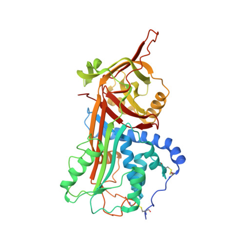
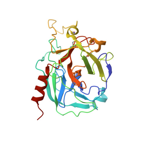
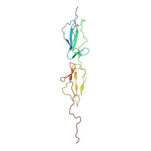
The Crystal Structure of the Michaelis-Menten Complex of C1 Esterase Inhibitor and C1s Reveals Novel Insights into Complement Regulation.

Garrigues, R.J.Garrison, M.P.Garcia, B.L.

(2024) J Immunol 

  • DOI: https://doi.org/10.4049/jimmunol.2400194
  • Primary Citation of Related Structures:  
    8W18

  • PubMed Abstract: 

    The ancient arm of innate immunity known as the complement system is a blood proteolytic cascade involving dozens of membrane-bound and solution-phase components. Although many of these components serve as regulatory molecules to facilitate controlled activation of the cascade, C1 esterase inhibitor (C1-INH) is the sole canonical complement regulator belonging to a superfamily of covalent inhibitors known as serine protease inhibitors (SERPINs). In addition to its namesake role in complement regulation, C1-INH also regulates proteases of the coagulation, fibrinolysis, and contact pathways. Despite this, the structural basis for C1-INH recognition of its target proteases has remained elusive. In this study, we present the crystal structure of the Michaelis-Menten (M-M) complex of the catalytic domain of complement component C1s and the SERPIN domain of C1-INH at a limiting resolution of 3.94 Å. Analysis of the structure revealed that nearly half of the protein/protein interface is formed by residues outside of the C1-INH reactive center loop. The contribution of these residues to the affinity of the M-M complex was validated by site-directed mutagenesis using surface plasmon resonance. Parallel analysis confirmed that C1-INH-interfacing residues on C1s surface loops distal from the active site also drive affinity of the M-M complex. Detailed structural comparisons revealed differences in substrate recognition by C1s compared with C1-INH recognition and highlight the importance of exosite interactions across broader SERPIN/protease systems. Collectively, this study improves our understanding of how C1-INH regulates the classical pathway of complement, and it sheds new light on how SERPINs recognize their cognate protease targets.


  • Organizational Affiliation

    Department of Microbiology and Immunology, Brody School of Medicine, East Carolina University, Greenville, NC.


Macromolecules
Find similar proteins by:  (by identity cutoff)  |  3D Structure
Entity ID: 1
MoleculeChains Sequence LengthOrganismDetailsImage
Plasma protease C1 inhibitorA [auth I]395Homo sapiensMutation(s): 0 
Gene Names: SERPING1C1INC1NH
UniProt & NIH Common Fund Data Resources
Find proteins for P05155 (Homo sapiens)
Explore P05155 
Go to UniProtKB:  P05155
PHAROS:  P05155
GTEx:  ENSG00000149131 
Entity Groups  
Sequence Clusters30% Identity50% Identity70% Identity90% Identity95% Identity100% Identity
UniProt GroupP05155
Sequence Annotations
Expand
  • Reference Sequence
Find similar proteins by:  (by identity cutoff)  |  3D Structure
Entity ID: 2
MoleculeChains Sequence LengthOrganismDetailsImage
Complement C1s subcomponent light chain251Homo sapiensMutation(s): 1 
Gene Names: C1S
UniProt & NIH Common Fund Data Resources
Find proteins for P09871 (Homo sapiens)
Explore P09871 
Go to UniProtKB:  P09871
PHAROS:  P09871
GTEx:  ENSG00000182326 
Entity Groups  
Sequence Clusters30% Identity50% Identity70% Identity90% Identity95% Identity100% Identity
UniProt GroupP09871
Sequence Annotations
Expand
  • Reference Sequence
Find similar proteins by:  (by identity cutoff)  |  3D Structure
Entity ID: 3
MoleculeChains Sequence LengthOrganismDetailsImage
Complement C1s subcomponent heavy chainC [auth A]151Homo sapiensMutation(s): 0 
Gene Names: C1S
UniProt & NIH Common Fund Data Resources
Find proteins for P09871 (Homo sapiens)
Explore P09871 
Go to UniProtKB:  P09871
PHAROS:  P09871
GTEx:  ENSG00000182326 
Entity Groups  
Sequence Clusters30% Identity50% Identity70% Identity90% Identity95% Identity100% Identity
UniProt GroupP09871
Sequence Annotations
Expand
  • Reference Sequence
Experimental Data & Validation

Experimental Data

  • Method: X-RAY DIFFRACTION
  • Resolution: 3.94 Å
  • R-Value Free: 0.281 
  • R-Value Work: 0.262 
  • R-Value Observed: 0.264 
  • Space Group: P 42 21 2
Unit Cell:
Length ( Å )Angle ( ˚ )
a = 168.628α = 90
b = 168.628β = 90
c = 88.342γ = 90
Software Package:
Software NamePurpose
PHENIXrefinement
XDSdata reduction
XDSdata scaling
PHENIXphasing

Structure Validation

View Full Validation Report


Entry History & Funding Information

Deposition Data


Funding OrganizationLocationGrant Number
National Institutes of Health/National Institute of General Medical Sciences (NIH/NIGMS)United States--

Revision History  (Full details and data files)

  • Version 1.0: 2024-06-26
    Type: Initial release
  • Version 1.1: 2024-07-24
    Changes: Author supporting evidence, Database references