8VME | pdb_00008vme

Crystal structure of the GSK-3/Axin complex bound to a phosphorylated beta-catenin T41A peptide


Experimental Data Snapshot

  • Method: X-RAY DIFFRACTION
  • Resolution: 2.30 Å
  • R-Value Free: 
    0.230 (Depositor), 0.230 (DCC) 
  • R-Value Work: 
    0.183 (DCC) 
  • R-Value Observed: 
    0.183 (Depositor) 

Starting Model: experimental
View more details

wwPDB Validation 3D Report Full Report

Validation slider image for 8VME

Ligand Structure Quality Assessment 


This is version 1.3 of the entry. See complete history

Literature

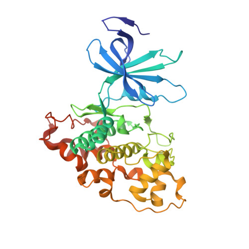
Structural and functional effects of phosphopriming and scaffolding in the kinase GSK-3 beta.

Enos, M.D.Gavagan, M.Jameson, N.Zalatan, J.G.Weis, W.I.

(2024) Sci Signal 17

  • DOI: https://doi.org/10.1126/scisignal.ado0881
  • Primary Citation Related Structures: 
    8VME, 8VMF, 8VMG

  • PubMed Abstract: 

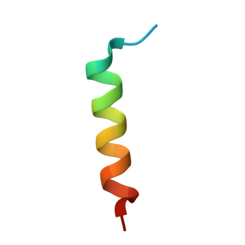
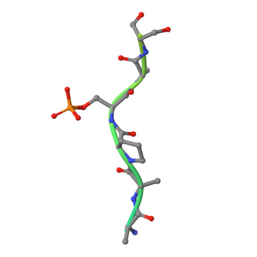
    Glycogen synthase kinase 3β (GSK-3β) targets specific signaling pathways in response to distinct upstream signals. We used structural and functional studies to dissect how an upstream phosphorylation step primes the Wnt signaling component β-catenin for phosphorylation by GSK-3β and how scaffolding interactions contribute to this reaction. Our crystal structure of GSK-3β bound to a phosphoprimed β-catenin peptide confirmed the expected binding mode of the phosphoprimed residue adjacent to the catalytic site. An aspartate phosphomimic in the priming site of β-catenin adopted an indistinguishable structure but reacted approximately 1000-fold slower than the native phosphoprimed substrate. This result suggests that substrate positioning alone is not sufficient for catalysis and that native phosphopriming interactions are necessary. We also obtained a structure of GSK-3β with an extended peptide from the scaffold protein Axin that bound with greater affinity than that of previously crystallized Axin fragments. This structure neither revealed additional contacts that produce the higher affinity nor explained how substrate interactions in the GSK-3β active site are modulated by remote Axin binding. Together, our findings suggest that phosphopriming and scaffolding produce small conformational changes or allosteric effects, not captured in the crystal structures, that activate GSK-3β and facilitate β-catenin phosphorylation. These results highlight limitations in our ability to predict catalytic activity from structure and have potential implications for the role of natural phosphomimic mutations in kinase regulation and phosphosite evolution.


  • Organizational Affiliation
    • Department of Structural Biology, Stanford University School of Medicine, Stanford, CA 94035, USA.

Macromolecule Content 

  • Total Structure Weight: 48.37 kDa 
  • Atom Count: 3,308 
  • Modeled Residue Count: 383 
  • Deposited Residue Count: 417 
  • Unique protein chains: 3

Macromolecules

Find similar proteins by:|  3D Structure
Entity ID: 1
MoleculeChains  Sequence LengthOrganismDetailsImage
Glycogen synthase kinase-3 beta365Mus musculusMutation(s): 0 
Gene Names: Gsk3b
EC: 2.7.11.26 (PDB Primary Data), 2.7.11.1 (PDB Primary Data)
UniProt
Find proteins for Q9WV60 (Mus musculus)
Explore Q9WV60 
Go to UniProtKB:  Q9WV60
Entity Groups
Sequence Clusters30% Identity50% Identity70% Identity90% Identity95% Identity100% Identity
UniProt GroupQ9WV60
Sequence Annotations
Expand
Reference Sequence
Find similar proteins by:  Sequence   |   3D Structure  
Entity ID: 2
MoleculeChains  Sequence LengthOrganismDetailsImage
Axin-124Homo sapiensMutation(s): 0 
Gene Names: Axin1AxinFu
UniProt & NIH Common Fund Data Resources
Find proteins for O15169 (Homo sapiens)
Explore O15169 
Go to UniProtKB:  O15169
PHAROS:  O15169
GTEx:  ENSG00000103126 
Entity Groups
Sequence Clusters30% Identity50% Identity70% Identity90% Identity95% Identity100% Identity
UniProt GroupO15169
Sequence Annotations
Expand
Reference Sequence
Find similar proteins by:|  3D Structure
Entity ID: 3
MoleculeChains  Sequence LengthOrganismDetailsImage
Catenin beta-128Homo sapiensMutation(s): 1 
Gene Names: CTNNB1CTNNBOK/SW-cl.35PRO2286
UniProt & NIH Common Fund Data Resources
Find proteins for P35222 (Homo sapiens)
Explore P35222 
Go to UniProtKB:  P35222
PHAROS:  P35222
GTEx:  ENSG00000168036 
Entity Groups
Sequence Clusters30% Identity50% Identity70% Identity90% Identity95% Identity100% Identity
UniProt GroupP35222
Sequence Annotations
Expand
Reference Sequence

Small Molecules

Ligands 4 Unique
IDChains Name / Formula / InChI Key2D Diagram3D Interactions
ADP

Query on ADP


Download:Ideal Coordinates CCD File
D [auth A]ADENOSINE-5'-DIPHOSPHATE
C10 H15 N5 O10 P2
XTWYTFMLZFPYCI-KQYNXXCUSA-N
GOL

Query on GOL


Download:Ideal Coordinates CCD File
G [auth A]
H [auth A]
I [auth A]
J [auth A]
K [auth A]
G [auth A],
H [auth A],
I [auth A],
J [auth A],
K [auth A],
L [auth A]
GLYCEROL
C3 H8 O3
PEDCQBHIVMGVHV-UHFFFAOYSA-N
MG

Query on MG


Download:Ideal Coordinates CCD File
E [auth A],
F [auth A]
MAGNESIUM ION
Mg
JLVVSXFLKOJNIY-UHFFFAOYSA-N
NA

Query on NA


Download:Ideal Coordinates CCD File
M [auth A]SODIUM ION
Na
FKNQFGJONOIPTF-UHFFFAOYSA-N
Modified Residues  1 Unique
IDChains TypeFormula2D DiagramParent
SEP
Query on SEP
C
L-PEPTIDE LINKINGC3 H8 N O6 PSER

Experimental Data & Validation

Experimental Data

  • Method: X-RAY DIFFRACTION
  • Resolution: 2.30 Å
  • R-Value Free:  0.230 (Depositor), 0.230 (DCC) 
  • R-Value Work:  0.183 (DCC) 
  • R-Value Observed: 0.183 (Depositor) 
Space Group: P 61 2 2
Unit Cell:
Length ( Å )Angle ( ˚ )
a = 82.257α = 90
b = 82.257β = 90
c = 280.99γ = 120
Software Package:
Software NamePurpose
Blu-Icedata collection
XDSdata reduction
Aimlessdata scaling
pointlessdata scaling
PHENIXphasing
PHENIXrefinement
Cootmodel building

Structure Validation

View Full Validation Report


Ligand Structure Quality Assessment 


Entry History 

& Funding Information

Deposition Data


Funding OrganizationLocationGrant Number
National Institutes of Health/National Institute of General Medical Sciences (NIH/NIGMS)United StatesGM131747
National Institutes of Health/National Institute of General Medical Sciences (NIH/NIGMS)United StatesGM119156
National Institutes of Health/National Institute of General Medical Sciences (NIH/NIGMS)United StatesT32 GM007276

Revision History  (Full details and data files)

  • Version 1.0: 2024-08-28
    Type: Initial release
  • Version 1.1: 2024-09-18
    Changes: Database references
  • Version 1.2: 2024-09-25
    Changes: Database references
  • Version 1.3: 2024-11-13
    Changes: Structure summary