Structural Comparison of Substrate Binding Sites in Dehaloperoxidase A and B.
Aktar, M.S., de Serrano, V., Ghiladi, R.A., Franzen, S.(2024) Biochemistry 63: 1761-1773
- PubMed: 38959050 Search on PubMed
- DOI: https://doi.org/10.1021/acs.biochem.4c00179
- Primary Citation Related Structures:
8VKC, 8VKD, 8VSK, 8VZR - PubMed Abstract:
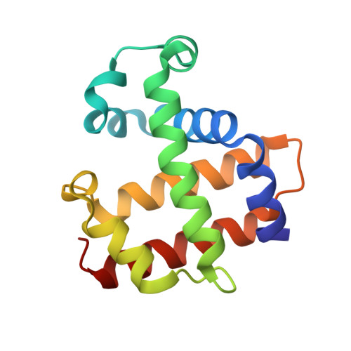
Dehalperoxidase (DHP) has diverse catalytic activities depending on the substrate binding conformation, pH, and dynamics in the distal pocket above the heme. According to our hypothesis, the molecular structure of the substrate and binding orientation in DHP guide enzymatic function. Enzyme kinetic studies have shown that the catalytic activity of DHP B is significantly higher than that of DHP A despite 96% sequence homology. There are more than 30 substrate-bound structures with DHP B, each providing insight into the nature of enzymatic binding at the active site. By contrast, the only X-ray crystallographic structures of small molecules in a complex with DHP A are phenols. This study is focused on investigating substrate binding in DHP A to compare with DHP B structures. Fifteen substrates were selected that were known to bind to DHP B in the crystal to test whether soaking substrates into DHP A would yield similar structures. Five of these substrates yielded X-ray crystal structures of substrate-bound DHP A, namely, 2,4-dichlorophenol (1.48 Å, PDB: 8EJN), 2,4-dibromophenol (1.52 Å, PDB: 8VSK), 4-nitrophenol (2.03 Å, PDB: 8VKC), 4-nitrocatechol (1.40 Å, PDB: 8VKD), and 4-bromo-o-cresol (1.64 Å, PDB: 8VZR). For the remaining substrates that bind to DHP B, such as cresols, 5-bromoindole, benzimidazole, 4,4-biphenol, 4.4-ethylidenebisphenol, 2,4-dimethoxyphenol, and guaiacol, the electron density maps in DHP A are not sufficient to determine the presence of the substrates, much less their orientation. In our hands, only phenols, 4-Br-o-cresol, and 4-nitrocatechol can be soaked into crystalline DHP A. None of the larger substrates were observed to bind. A minimum of seven hanging drops were selected for soaking with more than 50 crystals screened for each substrate. The five high-quality examples of direct comparison of modes of binding in DHP A and B for the same substrate provide further support for the hypothesis that the substrate-binding conformation determines the enzyme function of DHP.
- Department of Chemistry, North Carolina State University, Raleigh, North Carolina 27695, United States.
Organizational Affiliation: