8VK3

Structure of mouse RyR1 in complex with S100A1 (EGTA-only dataset)


Experimental Data Snapshot

  • Method: ELECTRON MICROSCOPY
  • Resolution: 3.21 Å
  • Aggregation State: PARTICLE 
  • Reconstruction Method: SINGLE PARTICLE 

wwPDB Validation   3D Report Full Report


This is version 1.1 of the entry. See complete history


Literature

Structural insights into the regulation of RyR1 by S100A1.

Weninger, G.Miotto, M.C.Tchagou, C.Reiken, S.Dridi, H.Brandenburg, S.Riedemann, G.C.Yuan, Q.Liu, Y.Chang, A.Wronska, A.Lehnart, S.E.Marks, A.R.

(2024) Proc Natl Acad Sci U S A 121: e2400497121-e2400497121

  • DOI: https://doi.org/10.1073/pnas.2400497121
  • Primary Citation of Related Structures:  
    8VJJ, 8VJK, 8VK3, 8VK4

  • PubMed Abstract: 

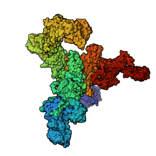
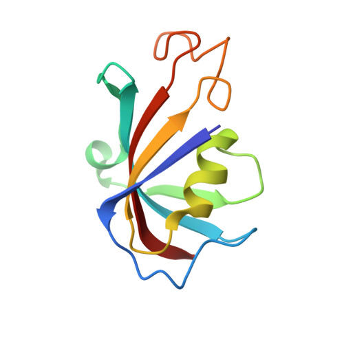
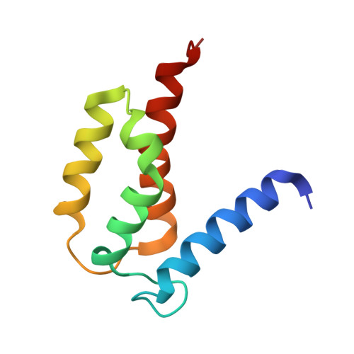
    S100A1, a small homodimeric EF-hand Ca 2+ -binding protein (~21 kDa), plays an important regulatory role in Ca 2+ signaling pathways involved in various biological functions including Ca 2+ cycling and contractile performance in skeletal and cardiac myocytes. One key target of the S100A1 interactome is the ryanodine receptor (RyR), a huge homotetrameric Ca 2+ release channel (~2.3 MDa) of the sarcoplasmic reticulum. Here, we report cryoelectron microscopy structures of S100A1 bound to RyR1, the skeletal muscle isoform, in absence and presence of Ca 2+ . Ca 2+ -free apo-S100A1 binds beneath the bridging solenoid (BSol) and forms contacts with the junctional solenoid and the shell-core linker of RyR1. Upon Ca 2+ -binding, S100A1 undergoes a conformational change resulting in the exposure of the hydrophobic pocket known to serve as a major interaction site of S100A1. Through interactions of the hydrophobic pocket with RyR1, Ca 2+ -bound S100A1 intrudes deeper into the RyR1 structure beneath BSol than the apo-form and induces sideways motions of the C-terminal BSol region toward the adjacent RyR1 protomer resulting in tighter interprotomer contacts. Interestingly, the second hydrophobic pocket of the S100A1-dimer is largely exposed at the hydrophilic surface making it prone to interactions with the local environment, suggesting that S100A1 could be involved in forming larger heterocomplexes of RyRs with other protein partners. Since S100A1 interactions stabilizing BSol are implicated in the regulation of RyR-mediated Ca 2+ release, the characterization of the S100A1 binding site conserved between RyR isoforms may provide the structural basis for the development of therapeutic strategies regarding treatments of RyR-related disorders.


  • Organizational Affiliation

    Department of Physiology and Cellular Biophysics, Center for Molecular Cardiology, Columbia University Vagelos College of Physicians and Surgeons, New York, NY 10032.


Macromolecules
Find similar proteins by:  (by identity cutoff)  |  3D Structure
Entity ID: 1
MoleculeChains Sequence LengthOrganismDetailsImage
Ryanodine receptor 1A,
N [auth D],
O [auth B],
P [auth C]
5,035Mus musculusMutation(s): 0 
UniProt & NIH Common Fund Data Resources
Find proteins for E9PZQ0 (Mus musculus)
Explore E9PZQ0 
Go to UniProtKB:  E9PZQ0
IMPC:  MGI:99659
Entity Groups  
Sequence Clusters30% Identity50% Identity70% Identity90% Identity95% Identity100% Identity
UniProt GroupE9PZQ0
Sequence Annotations
Expand
  • Reference Sequence
Find similar proteins by:  (by identity cutoff)  |  3D Structure
Entity ID: 2
MoleculeChains Sequence LengthOrganismDetailsImage
Peptidyl-prolyl cis-trans isomerase FKBP1AB [auth E],
E [auth H],
F,
G
108Mus musculusMutation(s): 0 
EC: 5.2.1.8
UniProt
Find proteins for P26883 (Mus musculus)
Explore P26883 
Go to UniProtKB:  P26883
Entity Groups  
Sequence Clusters30% Identity50% Identity70% Identity90% Identity95% Identity100% Identity
UniProt GroupP26883
Sequence Annotations
Expand
  • Reference Sequence
Find similar proteins by:  (by identity cutoff)  |  3D Structure
Entity ID: 3
MoleculeChains Sequence LengthOrganismDetailsImage
Protein S100A194Mus musculusMutation(s): 0 
Gene Names: S100a1
UniProt & NIH Common Fund Data Resources
Find proteins for Q91V77 (Mus musculus)
Explore Q91V77 
Go to UniProtKB:  Q91V77
IMPC:  MGI:1338917
Entity Groups  
Sequence Clusters30% Identity50% Identity70% Identity90% Identity95% Identity100% Identity
UniProt GroupQ91V77
Sequence Annotations
Expand
  • Reference Sequence
Small Molecules
Ligands 2 Unique
IDChains Name / Formula / InChI Key2D Diagram3D Interactions
PCW
Query on PCW

Download Ideal Coordinates CCD File 
BA [auth C]
R [auth A]
S [auth A]
T [auth D]
V [auth D]
BA [auth C],
R [auth A],
S [auth A],
T [auth D],
V [auth D],
X [auth B],
Y [auth B],
Z [auth C]
1,2-DIOLEOYL-SN-GLYCERO-3-PHOSPHOCHOLINE
C44 H85 N O8 P
SNKAWJBJQDLSFF-NVKMUCNASA-O
ZN (Subject of Investigation/LOI)
Query on ZN

Download Ideal Coordinates CCD File 
AA [auth C],
Q [auth A],
U [auth D],
W [auth B]
ZINC ION
Zn
PTFCDOFLOPIGGS-UHFFFAOYSA-N
Experimental Data & Validation

Experimental Data

  • Method: ELECTRON MICROSCOPY
  • Resolution: 3.21 Å
  • Aggregation State: PARTICLE 
  • Reconstruction Method: SINGLE PARTICLE 
EM Software:
TaskSoftware PackageVersion
MODEL REFINEMENTPHENIX
RECONSTRUCTIONcryoSPARC

Structure Validation

View Full Validation Report


Entry History & Funding Information

Deposition Data


Funding OrganizationLocationGrant Number
National Institutes of Health/National Heart, Lung, and Blood Institute (NIH/NHLBI)United StatesR01HL145473

Revision History  (Full details and data files)

  • Version 1.0: 2024-01-24
    Type: Initial release
  • Version 1.1: 2024-07-10
    Changes: Data collection, Database references