8VHL

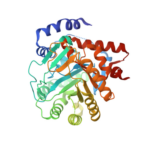
Structure of DHODH in Complex with Ligand 17


Experimental Data Snapshot

  • Method: X-RAY DIFFRACTION
  • Resolution: 1.93 Å
  • R-Value Free: 0.199 
  • R-Value Work: 0.168 
  • R-Value Observed: 0.169 

Starting Model: experimental
View more details

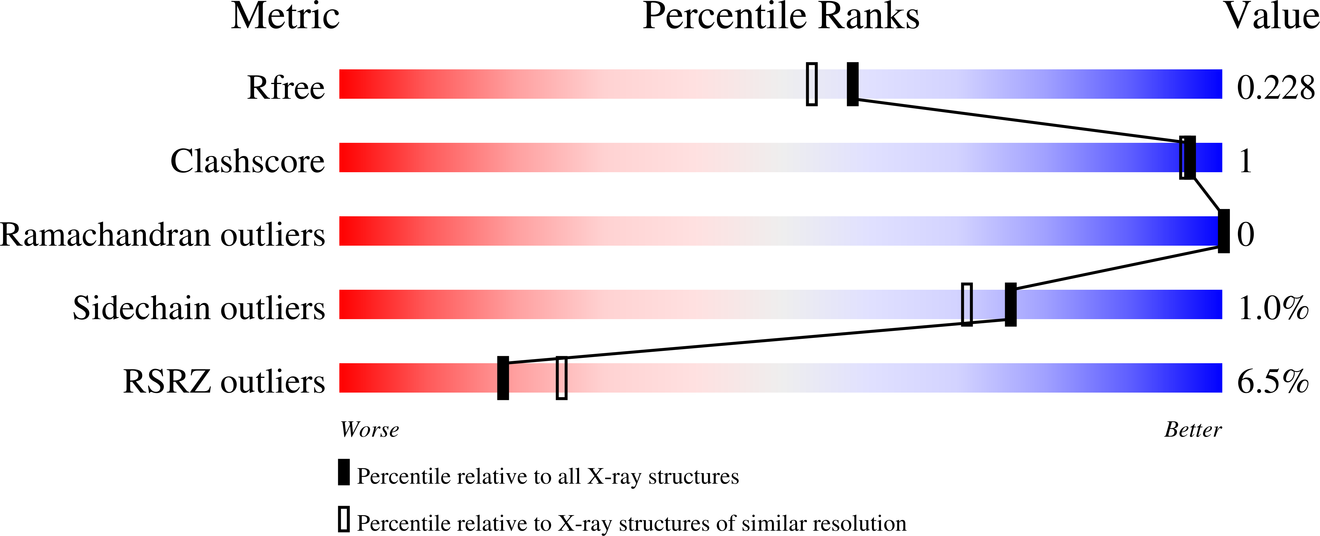
wwPDB Validation   3D Report Full Report


Ligand Structure Quality Assessment 


This is version 1.0 of the entry. See complete history


Literature

Discovery of Alternative Binding Poses through Fragment-Based Identification of DHODH Inhibitors.

DeRatt, L.G.Pietsch, E.C.Cisar, J.S.Jacoby, E.Kazmi, F.Matico, R.Shaffer, P.Tanner, A.Wang, W.Attar, R.Edwards, J.P.Kuduk, S.D.

(2024) ACS Med Chem Lett 15: 381-387

  • DOI: https://doi.org/10.1021/acsmedchemlett.3c00543
  • Primary Citation of Related Structures:  
    8VHL, 8VHM

  • PubMed Abstract: 

    Dihydroorotate dehydrogenase (DHODH) is a mitochondrial enzyme that affects many aspects essential to cell proliferation and survival. Recently, DHODH has been identified as a potential target for acute myeloid leukemia therapy. Herein, we describe the identification of potent DHODH inhibitors through a scaffold hopping approach emanating from a fragment screen followed by structure-based drug design to further improve the overall profile and reveal an unexpected novel binding mode. Additionally, these compounds had low P-gp efflux ratios, allowing for applications where exposure to the brain would be required.


  • Organizational Affiliation

    Janssen Pharmaceutical Research and Development, 1400 McKean Rd., Spring House, Pennsylvania 19477, United States.


Macromolecules
Find similar proteins by:  (by identity cutoff)  |  3D Structure
Entity ID: 1
MoleculeChains Sequence LengthOrganismDetailsImage
Dihydroorotate dehydrogenase (quinone), mitochondrial369Homo sapiensMutation(s): 0 
Gene Names: DHODH
EC: 1.3.5.2
UniProt & NIH Common Fund Data Resources
Find proteins for Q02127 (Homo sapiens)
Explore Q02127 
Go to UniProtKB:  Q02127
GTEx:  ENSG00000102967 
Entity Groups  
Sequence Clusters30% Identity50% Identity70% Identity90% Identity95% Identity100% Identity
UniProt GroupQ02127
Sequence Annotations
Expand
  • Reference Sequence
Small Molecules
Ligands 6 Unique
IDChains Name / Formula / InChI Key2D Diagram3D Interactions
A1AA2 (Subject of Investigation/LOI)
Query on A1AA2

Download Ideal Coordinates CCD File 
D [auth A]N-(2-chloro-6-fluorophenyl)-4-[ethyl(2-hydroxyethyl)carbamamido]-5-fluoro-2-{[(2S)-1,1,1-trifluoropropan-2-yl]oxy}benzamide
C21 H21 Cl F5 N3 O4
LBIUCQQLEKERKJ-NSHDSACASA-N
FMN
Query on FMN

Download Ideal Coordinates CCD File 
B [auth A]FLAVIN MONONUCLEOTIDE
C17 H21 N4 O9 P
FVTCRASFADXXNN-SCRDCRAPSA-N
ORO
Query on ORO

Download Ideal Coordinates CCD File 
C [auth A]OROTIC ACID
C5 H4 N2 O4
PXQPEWDEAKTCGB-UHFFFAOYSA-N
SO4
Query on SO4

Download Ideal Coordinates CCD File 
G [auth A],
K [auth A],
L [auth A]
SULFATE ION
O4 S
QAOWNCQODCNURD-UHFFFAOYSA-L
GOL
Query on GOL

Download Ideal Coordinates CCD File 
I [auth A],
J [auth A],
M [auth A],
N [auth A]
GLYCEROL
C3 H8 O3
PEDCQBHIVMGVHV-UHFFFAOYSA-N
ACT
Query on ACT

Download Ideal Coordinates CCD File 
E [auth A],
F [auth A],
H [auth A]
ACETATE ION
C2 H3 O2
QTBSBXVTEAMEQO-UHFFFAOYSA-M
Experimental Data & Validation

Experimental Data

  • Method: X-RAY DIFFRACTION
  • Resolution: 1.93 Å
  • R-Value Free: 0.199 
  • R-Value Work: 0.168 
  • R-Value Observed: 0.169 
  • Space Group: P 32 2 1
Unit Cell:
Length ( Å )Angle ( ˚ )
a = 90.744α = 90
b = 90.744β = 90
c = 122.713γ = 120
Software Package:
Software NamePurpose
XDSdata reduction
XSCALEdata scaling
REFMACrefinement
PHASERphasing

Structure Validation

View Full Validation Report


Ligand Structure Quality Assessment 


Entry History & Funding Information

Deposition Data


Funding OrganizationLocationGrant Number
Not funded--

Revision History  (Full details and data files)

  • Version 1.0: 2024-04-03
    Type: Initial release