8UH9 | pdb_00008uh9

Crystal structure of SARS-CoV-2 main protease E166V mutant in complex with an inhibitor TKB-272


Experimental Data Snapshot

  • Method: X-RAY DIFFRACTION
  • Resolution: 2.07 Å
  • R-Value Free: 
    0.240 (Depositor), 0.248 (DCC) 
  • R-Value Work: 
    0.192 (Depositor), 0.182 (DCC) 
  • R-Value Observed: 
    0.194 (Depositor) 

Starting Model: experimental
View more details

wwPDB Validation 3D Report Full Report

Validation slider image for 8UH9

Ligand Structure Quality Assessment 


This is version 1.2 of the entry. See complete history

Literature

An orally available P1'-5-fluorinated M pro inhibitor blocks SARS-CoV-2 replication without booster and exhibits high genetic barrier.

Higashi-Kuwata, N.Bulut, H.Hayashi, H.Tsuji, K.Ogata-Aoki, H.Kiso, M.Takamune, N.Kishimoto, N.Hattori, S.I.Ishii, T.Kobayakawa, T.Nakano, K.Shimizu, Y.Das, D.Saruwatari, J.Hasegawa, K.Murayama, K.Sukenaga, Y.Takamatsu, Y.Yoshimura, K.Aoki, M.Furusawa, Y.Okamura, T.Yamayoshi, S.Kawaoka, Y.Misumi, S.Tamamura, H.Mitsuya, H.

(2025) PNAS Nexus 4: pgae578-pgae578

  • DOI: https://doi.org/10.1093/pnasnexus/pgae578
  • Primary Citation Related Structures: 
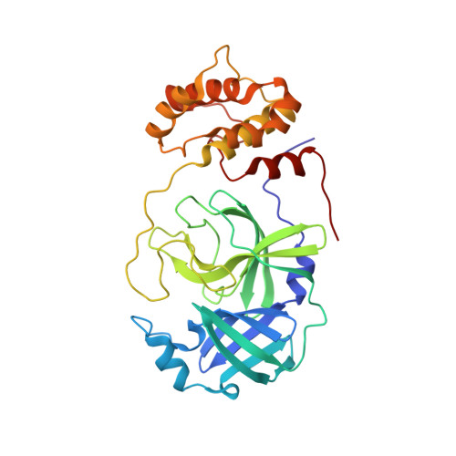
    8UH5, 8UH8, 8UH9

  • PubMed Abstract: 

    We identified a 5-fluoro-benzothiazole-containing small molecule, TKB272, through fluorine-scanning of the benzothiazole moiety, which more potently inhibits the enzymatic activity of SARS-CoV-2's main protease (M pro ) and more effectively blocks the infectivity and replication of all SARS-CoV-2 strains examined including Omicron variants such as SARS-CoV-2 XBB1.5 and SARS-CoV-2 EG.5.1 than two M pro inhibitors: nirmatrelvir and ensitrelvir. Notably, the administration of ritonavir-boosted nirmatrelvir and ensitrelvir causes drug-drug interactions warranting cautions due to their CYP3A4 inhibition, thereby limiting their clinical utility. When orally administered, TKB272 blocked SARS-CoV-2 XBB1.5 replication without ritonavir in B6.Cg-Tg(K18-hACE2)2-Prlmn/J-transgenic mice, comparably as did ritonavir-boosted nirmatrelvir. When the ancestral SARS-CoV-2 was propagated with nirmatrelvir in vitro, a highly nirmatrelvir-resistant E166V-carrying variant (SARS-CoV-2 E166V-P14 ) readily emerged by passage 14; however, when propagated with TKB272, no variants emerged by passage 25. SARS-CoV-2 E166V showed some cross-resistance to TKB272 but was substantially sensitive to the compound. X-ray structural analyses and mass-spectrometric data showed that the E166V substitution disrupts the critical dimerization-initiating Ser1'-E166 interactions, thereby limiting nirmatrelvir's M pro inhibition but that TKB272 nevertheless forms a tight binding with M pro 's catalytic active sight even in the presence of the E166V substitution. TKB272 shows no apparent genotoxicity as tested in the micro-Ames test. Highly potent TKB272 may serve as a COVID-19 therapeutic, overcome resistance to existing M pro inhibitors.


  • Organizational Affiliation
    • Department of Refractory Viral Diseases, National Center for Global Health and Medicine Research Institute, 1-21-1 Toyama, Shinjuku-ku, Tokyo 162-8655, Japan.

Macromolecule Content 

  • Total Structure Weight: 34.56 kDa 
  • Atom Count: 2,421 
  • Modeled Residue Count: 306 
  • Deposited Residue Count: 306 
  • Unique protein chains: 1

Macromolecules

Find similar proteins by:|  3D Structure
Entity ID: 1
MoleculeChains  Sequence LengthOrganismDetailsImage
3C-like proteinase nsp5306Severe acute respiratory syndrome coronavirus 2Mutation(s): 1 
Gene Names: rep1a-1b
EC: 3.4.22.69
UniProt
Find proteins for P0DTD1 (Severe acute respiratory syndrome coronavirus 2)
Explore P0DTD1 
Go to UniProtKB:  P0DTD1
Entity Groups
Sequence Clusters30% Identity50% Identity70% Identity90% Identity95% Identity100% Identity
UniProt GroupP0DTD1
Sequence Annotations
Expand
Reference Sequence

Small Molecules

Ligands 2 Unique
IDChains Name / Formula / InChI Key2D Diagram3D Interactions
WOK
(Subject of Investigation/LOI)

Query on WOK


Download:Ideal Coordinates CCD File
B [auth A](1R,2S,5S)-N-{(1S,2S)-1-(5-fluoro-1,3-benzothiazol-2-yl)-1-hydroxy-3-[(3S)-2-oxopyrrolidin-3-yl]propan-2-yl}-6,6-dimethyl-3-[3-methyl-N-(trifluoroacetyl)-L-valyl]-3-azabicyclo[3.1.0]hexane-2-carboxamide
C30 H37 F4 N5 O5 S
AVAISVNJDSSFQV-UDQBHXPNSA-N
PEG

Query on PEG


Download:Ideal Coordinates CCD File
C [auth A]DI(HYDROXYETHYL)ETHER
C4 H10 O3
MTHSVFCYNBDYFN-UHFFFAOYSA-N

Experimental Data & Validation

Experimental Data

  • Method: X-RAY DIFFRACTION
  • Resolution: 2.07 Å
  • R-Value Free:  0.240 (Depositor), 0.248 (DCC) 
  • R-Value Work:  0.192 (Depositor), 0.182 (DCC) 
  • R-Value Observed: 0.194 (Depositor) 
Space Group: C 1 2 1
Unit Cell:
Length ( Å )Angle ( ˚ )
a = 113.673α = 90
b = 53.09β = 101.96
c = 45.774γ = 90
Software Package:
Software NamePurpose
REFMACrefinement
DIALSdata reduction
DIALSdata scaling
MOLREPphasing

Structure Validation

View Full Validation Report


Ligand Structure Quality Assessment 


Entry History 

& Funding Information

Deposition Data


Funding OrganizationLocationGrant Number
National Institutes of Health/National Cancer Institute (NIH/NCI)United States--

Revision History  (Full details and data files)

  • Version 1.0: 2023-12-20
    Type: Initial release
  • Version 1.1: 2024-10-23
    Changes: Structure summary
  • Version 1.2: 2025-02-19
    Changes: Database references