8TMV | pdb_00008tmv

HTLV-1 capsid protein N-terminal domain triclinic crystal form with phosphate ion


Experimental Data Snapshot

  • Method: X-RAY DIFFRACTION
  • Resolution: 1.73 Å
  • R-Value Free: 
    0.230 (Depositor), 0.230 (DCC) 
  • R-Value Work: 
    0.200 (Depositor), 0.200 (DCC) 
  • R-Value Observed: 
    0.202 (Depositor) 

Starting Model: experimental
View more details

wwPDB Validation 3D Report Full Report

Validation slider image for 8TMV

Ligand Structure Quality Assessment 


This is version 1.2 of the entry. See complete history

Literature

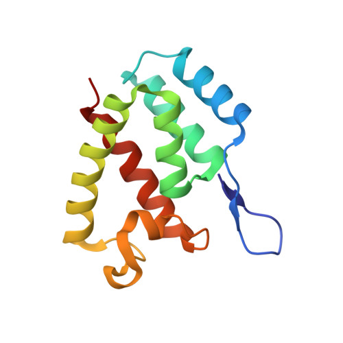
The Human T-cell Leukemia Virus capsid protein is a potential drug target.

Yu, R.Phalora, P.Li, N.Bocking, T.Jacques, D.A.

(2025) Nat Commun 16: 10892-10892

  • DOI: https://doi.org/10.1038/s41467-025-65899-2
  • Primary Citation Related Structures: 
    8ERE, 8ERF, 8ERG, 8ERH, 8ERI, 8TMV, 8TMW

  • PubMed Abstract: 

    Human T-cell Leukaemia Virus type 1 (HTLV-1) is an untreatable retrovirus that causes lethal malignancies and degenerative inflammatory conditions. Effective treatments have been delayed by substantial gaps in our knowledge of the fundamental virology, especially when compared to the closely related virus, HIV. A recently developed and highly effective anti-HIV strategy is to target the virus with drugs that interfere with capsid integrity and interactions with the host. Importantly, the first in-class anti-capsid drug approved, lenacapavir, can provide long-acting pre-exposure prophylaxis. Such a property would provide a means to prevent the transmission of HTLV-1, but its capsid has not previously been considered as a drug target. Here we describe high-resolution crystal structures of the HTLV-1 capsid protein, define essential lattice interfaces, and identify a distinct ligand-binding pocket. We show that this pocket is essential for virus infectivity, providing a potential target for future anti-capsid drug development.


  • Organizational Affiliation
    • Department of Molecular Medicine, School of Biomedical Sciences, University of New South Wales, Sydney, NSW, Australia.

Macromolecule Content 

  • Total Structure Weight: 14.24 kDa 
  • Atom Count: 1,049 
  • Modeled Residue Count: 125 
  • Deposited Residue Count: 127 
  • Unique protein chains: 1

Macromolecules

Find similar proteins by:|  3D Structure
Entity ID: 1
MoleculeChains  Sequence LengthOrganismDetailsImage
capsid protein p24127HTLV-1 subtype CMutation(s): 0 
UniProt
Find proteins for U3RC00 (Human T-cell leukemia virus type I)
Explore U3RC00 
Go to UniProtKB:  U3RC00
Entity Groups
Sequence Clusters30% Identity50% Identity70% Identity90% Identity95% Identity100% Identity
UniProt GroupU3RC00
Sequence Annotations
Expand
Reference Sequence

Small Molecules

Ligands 1 Unique
IDChains Name / Formula / InChI Key2D Diagram3D Interactions
PO4
(Subject of Investigation/LOI)

Query on PO4


Download:Ideal Coordinates CCD File
B [auth A]PHOSPHATE ION
O4 P
NBIIXXVUZAFLBC-UHFFFAOYSA-K

Experimental Data & Validation

Experimental Data

  • Method: X-RAY DIFFRACTION
  • Resolution: 1.73 Å
  • R-Value Free:  0.230 (Depositor), 0.230 (DCC) 
  • R-Value Work:  0.200 (Depositor), 0.200 (DCC) 
  • R-Value Observed: 0.202 (Depositor) 
Space Group: P 1
Unit Cell:
Length ( Å )Angle ( ˚ )
a = 28.195α = 65.19
b = 30.72β = 79.3
c = 36.081γ = 89.56
Software Package:
Software NamePurpose
PHENIXrefinement
Aimlessdata scaling
PHASERphasing
XDSdata reduction

Structure Validation

View Full Validation Report


Ligand Structure Quality Assessment 


Entry History 

& Funding Information

Deposition Data


Funding OrganizationLocationGrant Number
Wellcome TrustUnited Kingdom214344/Z/18/Z

Revision History  (Full details and data files)

  • Version 1.0: 2024-08-07
    Type: Initial release
  • Version 1.1: 2025-12-17
    Changes: Database references, Structure summary
  • Version 1.2: 2026-03-04
    Changes: Refinement description